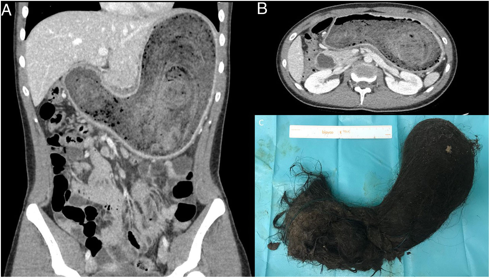
A 17-year-old female patient consulted for nausea, vomiting and bilious vomiting, with no abdominal pain. On examination, a palpable mass was observed in the epigastrium. The CT scan revealed a large, organized, concentric mass that occupied the entire gastric cavity (12 201 cc), suggestive of a trichobezoar (Fig. 1). Initially, endoscopic treatment was attempted, which was impossible, so a transverse laparotomy was ultimately performed. The specimen was extracted by means of a gastrotomy along the greater curvature (C). In the postoperative period, the patient presented infection of the surgical wound and an intra-abdominal collection, which was treated with ultrasound-guided percutaneous drainage. The patient progressed favorably and was discharged with a referral to the department of psychiatry for evaluation.
FinancingThis study was not funded by research grants.
Please cite this article as: Villalabeitia Ateca I, Alonso Calderón E, Alonso Carnicero P, Errazti Olartekoetxea G. Tricobezoar gástrico gigante en una paciente adolescente. Cir Esp. 2021;99:65.


