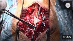
Material y método. Estudio experimental de 67 ratas sometidas a traumatismo esplénico quirúrgico controlado divididas en dos grupos: grupo A, esplenectomía parcial polar inferior y grupo B, laceraciones capsulares esplénicas. Se estudiaron 4 subgrupos, diferenciados entre sí en función del tiempo transcurrido tras la intervención: 48 h, 14 días y 30 y 90 días, valorándose los hallazgos macroscópicos e histológicos encontrados tras su sacrificio.
Resultados. Hubo 2 fallecimientos por hemoperitoneo masivo y un fallecimiento por evisceración (4,5%) entre los animales de experimentación. En el resto de las ratas la lesión cicatrizó sin problemas, destacando en los primeros momentos el papel ejercido por las adherencias del epiplón y órganos vecinos para cohibir la hemorragia. A largo plazo, la formación de una "neocápsula" de mayor grosor que la habitual del bazo, a expensas de un tejido de granulación, constituido por vasos neoformados e infiltrados inflamatorios, fue el factor determinante de la correcta cicatrización posterior.
Conclusiones. La cicatrización espontánea de las lesiones esplénicas es un proceso perfectamente plausible y seguro en el animal de experimentación utilizado. Con las reservas propias de la extrapolación del modelo experimental al humano, este estudio también podría sugerir que la esplenectomía por traumatismo debería comenzar a considerarse "de necesidad" y no "de principio".
Material and method. An experimental study was carried out in 67 rats in which splenic trauma was provoked under controlled surgical conditions. The animals were divided into two groups: group A, which was subjected to partial splenectomy involving the lower pole; and group B, subjected to splenic capsular laceration. These two groups were further divided into four subgroups, differentiated with respect to the postoperative time elapsed: 48 hours and 14, 30 and 90 days. A subgroup from each group of rats was sacrificed at each of these times and the macroscopic and histological findings assessed.
Results. Two animals died of massive hemoperitoneum and one of evisceration (4.5%). In the remainder of the rats, the injuries healed without problems. Moreover, in the initial moments, adhesions involving omentum and other neighboring organs played an important role in limiting hemorrhage. Over the long term, the acquisition by the spleen of a thicker "neocapsule" than before, comprised of newly formed vessels and inflammatory infiltrates at the expense of a granulation tissue, was decisive in achieving proper healing.
Conclusions. Spontaneous healing of splenic lesions is a perfectly feasible and safe process in the experimental animal employed in the present study. Cautiously extrapolating the experimental model to the human, the results suggest that options other than splenectomy should be considered in the management of splenic trauma


