


Pleural empyema is an infrequent manifestation of extraintestinal Clostridioidesdifficile infection, with just eight cases reported in literature.
MethodsWe report a new case in a 70-year-old male without comorbidities or evidence of concomitant gastrointestinal disease, and review the previous cases reported in the literature.
ResultsThe isolate was susceptible to all antimicrobial tested and was negative for A+B toxins. The patient fully recovered after drainages and antimicrobial therapy with amoxicillin–clavulanate and doxycycline.
ConclusionAs in the previously reported cases, aspiration was the most plausible hypothesis of mechanism of infection in our patient. Empyema by Clostridioidesdifficile is a diagnostic challenge, since it is necessary to rule out that the isolation of this microorganism in pleural fluid is not a contamination. Furthermore, more evidence is needed for its treatment since data regarding this entity are still scarce.
El empiema pleural es una manifestación infrecuente de la infección extraintestinal por Clostridioidesdifficile, con sólo ocho casos reportados en la literatura.
MétodosDocumentamos un nuevo caso de un varón de 70 años sin comorbilidades ni evidencia de enfermedad gastrointestinal concomitante y revisamos los casos previamente descritos en la literatura.
ResultadosEl aislado fue sensible a todos los antibióticos testados y fue negativo para las toxinas A+B. El paciente se recuperó totalmente tras la realización de drenajes y terapia antimicrobiana con amoxicilina-clavulánico y doxiciclina.
ConclusiónAl igual que en los casos previamente documentados, la broncoaspiración fue la hipótesis más plausible del mecanismo de infección en nuestro paciente. El empiema por Clostridioidesdifficile constituye un reto diagnóstico, ya que es necesario descartar que su aislamiento en líquido pleural no se deba a una contaminación. Además, se necesita más evidencia científica para el tratamiento de esta entidad, ya que los datos sobre la misma aún son escasos.
Clostridioides difficile is a Gram-positive, anaerobe spore forming rod that can form part of the gastrointestinal commensal microbiota in up to 17.5% of healthy adults.1 Firstly identified in 1935, it was originally named Bacillus difficilis because of its difficulty to be isolated in the laboratory.2 It is especially well known because some strains are producers of toxins A and B and cause antibiotic-associated diarrhea, whose severity ranges from a self-limiting infection to a life-threatening pseudomembranous colitis.2
Extracolonic C. difficile infection (CDI) is rarely reported and commonly associated with abdominal surgery. Moreover, isolation of C. difficile from extraintestinal locations, found as part of polimicrobial microbiota, is often difficult to interpret and commonly considered as contamination.3,4 Pleural empyema is a very infrequent manifestation of extraintestinal CDI, with just eight cases reported in literature.5–11
Here we present a rare case of pleural empyema caused by C. difficile, acquired in the community, in a patient without evidence of concomitant gastrointestinal disease. Also, the literature available of this entity is reviewed.
Case reportA 70-year-old male presented to our hospital, a 1000-bed tertiary hospital in northern Spain, with a one-day history of right-sided pleuritic chest pain. He did not report fever, shortness of breath or diarrhea. The weeks before admission, he had received amoxicillin as prophylaxis before a dental extraction. Significant past medical history included complicated diverticulitis and bacteremia by Eggerthellalenta.
Physical examination showed diminished breath sounds in the right lower lobe. Blood analysis demonstrated a leukocyte count of 29.96×10⁹/l (4.5–10×109/l) and a C reactive protein of 202mg/l (0–5mg/l).
A chest radiograph and computed tomography (CT) revealed a right-sided pleural effusion and atelectasis of the lower lobe of the right lung. There were no pathological findings in upper quadrants of abdomen. Analysis of pleural fluid, obtained after the insertion of a 14-Fr pigtail catheter in the pleural cavity, was suggestive of empyema (pH 6.8, glucose 2mg/dl, lactate dehydrogenase 7591U/l). Empirical treatment with amoxicillin–clavulanate 1000mg/200mg iv q8 hours was started and intrapleural fibrinolytics were administrated, with good clinical and radiological outcome. Cultures of pleural fluid, performed following the Infectious Diseases Society of America (IDSA) recommendations, were positive for two different bacteria identified by MALDI TOF/MS (Bruker Daltonics, Bremen, Germany) as Staphylococcus hominis and C. difficile, which were initially interpreted as contaminants. Antibiogram was performed by Etest strips (bioMérieux, Marcy l’Etoile, France) and interpreted according to 2022 EUCAST C. difficile breakpoints (https://www.eucast.org/clinical_breakpoints) for vancomycin and metronidazole; and CLSI breakpoints for anaerobes12 for tetracycline, amoxicillin/clavulanate and clindamycin. The isolate was susceptible to all the antimicrobials tested. The patient was discharged home eight days after admission with oral antibiotic therapy (amoxicillin–clavulanate 875/125mg po BID 10 days).
A month and a half later, the patient was readmitted due to worsening general condition. He referred a growing mass in upper right lumbar region, but continued afebrile. A chest CT was performed, showing a multiloculated collection of 4.2cm×7.7cm×6.3cm in the right pleural cavity which extended across the diaphragm into the right retroperitoneal space (5.1cm×7cm×14cm) and infiltrated lumbar paravertebral muscles. Antibiotic therapy with amoxicillin–clavulanate was reinitiated and an 8F catheter was inserted in the retroperitoneal collection, with removal of 250 cm3 of purulent fluid. Radiological improvement was observed in CT control and subsequent pus cultures were positive for C. difficile, with no other species recovered. Despite the lack of gastrointestinal symptoms, a stool sample was collected, and the FilmArray Gastrointestinal PCR Panel (BioFire Diagnostics, Salt Lake City, UT, USA) was performed, being positive for C. difficile but negative for A+B toxin, as well as the isolate recovered from the collection. The patient was readmitted two weeks after his second discharge due to gastrointestinal side effects of amoxicillin–clavulanate, a new retroperitoneal drainage was performed, and then he completed six weeks of oral treatment with doxycycline (100mg PO q12 hours) with complete recovery.
DiscussionC. difficile is a well-established cause of antibiotic-associated diarrhea. It was firstly identified in 1935, among the normal gastrointestinal microbiota in infants, although its pathogenic potential would not be recognized until the 1970s.2 Although it has been traditionally considered a health-care associated pathogen, there has been an increase in its community incidence in the last years.4
Extraintestinal CDI are infrequent, and tend to appear in hospitalized patients with comorbidities. Many of the cases described in literature occur after abdominal surgery, with the development of intra-abdominal abscesses, peritonitis or abdominal wound infections.4 Brain abscesses, osteomyelitis, reactive arthritis, pyelonephritis, pleural empyema, soft-tissue infection, appendicitis, pericarditis and prosthetic joint infection have also been reported.2,4 These manifestations, not always caused by toxigenic isolates, frequently happen without a recent history of diarrhea, suggesting that their low intestinal virulence allows prolonged carriage which is followed by opportunistic infections.2 Bacteremia by C. difficile, although rarely described, could be implicated as a route of transmission of extraintestinal CDI.3,4
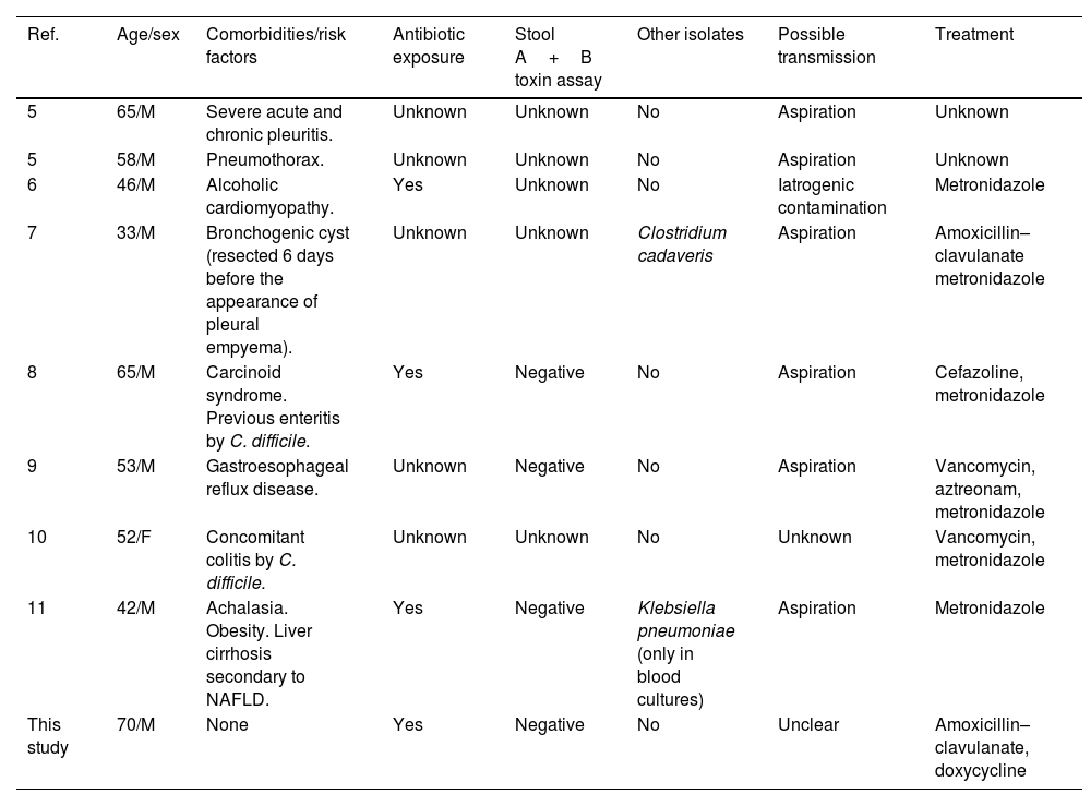
There are only a few cases of pleural empyema reported in literature (Table 1).5–11 Smith and King described the two first in 1962, in a series of patients with various extraintestinal CDI. Both cases were attributed to aspiration,5 which was also the most likely mechanism of infection in most of the other cases described. Patients from other reports had a history of achalasia, gastroesophageal reflux disease or had been previously treated with proton pump inhibitors, as predisposing factors.9,11 Iatrogenic contamination of pleural space was another hypothesis raised as a source of infection, since in two reports two patients had undergone prior thoracocentesis.6,11 Hematogenous spread as mechanism of infection was never described.
Clinical features of patients with pleural empyema caused by Clostridioides difficile.
| Ref. | Age/sex | Comorbidities/risk factors | Antibiotic exposure | Stool A+B toxin assay | Other isolates | Possible transmission | Treatment |
|---|---|---|---|---|---|---|---|
| 5 | 65/M | Severe acute and chronic pleuritis. | Unknown | Unknown | No | Aspiration | Unknown |
| 5 | 58/M | Pneumothorax. | Unknown | Unknown | No | Aspiration | Unknown |
| 6 | 46/M | Alcoholic cardiomyopathy. | Yes | Unknown | No | Iatrogenic contamination | Metronidazole |
| 7 | 33/M | Bronchogenic cyst (resected 6 days before the appearance of pleural empyema). | Unknown | Unknown | Clostridium cadaveris | Aspiration | Amoxicillin–clavulanate metronidazole |
| 8 | 65/M | Carcinoid syndrome. Previous enteritis by C. difficile. | Yes | Negative | No | Aspiration | Cefazoline, metronidazole |
| 9 | 53/M | Gastroesophageal reflux disease. | Unknown | Negative | No | Aspiration | Vancomycin, aztreonam, metronidazole |
| 10 | 52/F | Concomitant colitis by C. difficile. | Unknown | Unknown | No | Unknown | Vancomycin, metronidazole |
| 11 | 42/M | Achalasia. Obesity. Liver cirrhosis secondary to NAFLD. | Yes | Negative | Klebsiella pneumoniae (only in blood cultures) | Aspiration | Metronidazole |
| This study | 70/M | None | Yes | Negative | No | Unclear | Amoxicillin–clavulanate, doxycycline |
M, male; F, female; NAFLD, nonalcoholic fatty liver disease.
As occurred in our patient, previous antibiotic exposure was described in three of the previous reported cases (in the remaining five, this data was not specified). Stool toxin assay was negative in three cases. In the remaining five, this test was not performed or data is unclear. However, one of the patients with negative stool toxin assay had a medical history of C. difficile colitis,8 and another presented indicative signs of concurrent C. difficile colitis in a CT of abdomen performed after hospital admission.10 Our patient did not have symptoms of concomitant gastrointestinal disease, nor radiological findings suggestive of colitis, furthermore, his stool sample revealed the presence of a non-toxigenic C. difficile strain. The mechanism of infection remains unclear in the case here reported. The patient could have experienced a transient bacteremia with bacterial migration in the pleural space, although this possibility has never been described in literature. In fact, in a review of C. difficile bacteremia by Libby and Bearman, hematogenous spread was not described as a mechanism of pleuropulmonary infection.4 The most plausible hypothesis would be bronchial aspiration of C. difficile spores, subsequent germination and pneumonia development with pleural exudative effusion. C. difficile can be isolated from oral cavity in hospitalized patients, although is not part of the normal microbiota of this area.8 Cultures of the oral cavity were not performed in our patient nor in any of the other cases of pleural empyema, except in the one reported by Hudson et al., in which it was negative.8
The approach of extra-intestinal CDI treatment is controversial, since antibiotic regimens are not well defined neither in guidelines or in the literature. Previous described cases of empyema by C. difficile were treated with intravenous metronidazole or vancomycin, as well as beta-lactam antibiotics such as amoxicillin–clavulanate or cefazoline. In six of the cases previously reported the patients experienced full recovery (in the remaining two cases, this information is not available).5–11 Our patient was initially treated with amoxicillin–clavulanate, and completed six weeks of therapy with oral doxycycline, due to gastrointestinal side effects associated to the first antibiotic, with full recovery. Tetracyclines may serve as an alternative or a component of combination therapy for CDI associated diarrhea, and most isolates reported in Europe are susceptible, with resistance rates under 10%. Doxycycline may prevent or attenuate the virulence factors of toxigenic C. difficile.13 Also, this drug has many advantages over other tetracyclines, including less-frequent dosing, improved absorption, greater tissue distribution, longer half-life, and less photosensitivity.14
In conclusion, pleural empyema is a rare manifestation of extraintestinal Clostridioidesdifficile infection, with just eight cases reported in literature and is a diagnostic challenge, since it is necessary to rule out that the isolation of this microorganism in pleural fluid is not a contamination. Furthermore, more evidence is needed for the treatment of extraintestinal CDI in general, and in empyema in particular since data regarding the treatment of this entity is still scarce.
FundingThis work received no specific grant from any funding agency in the public, commercial.
Conflicts of interestThe authors declare no conflicts of interest, regarding this work.

