OVERVIEW
Helicobacter pylori eradication is crucial in the treatment of peptic ulcer disease, and plays a key role in modifying the natural course of the disease1,2. However, the benefits of H. pylori on symptom control is controversial, specially in patients with non-ulcer dyspepsia3. The reduction in ulcer recurrence observed following eradication of the bacteria improves symptoms and quality of life of peptic ulcer patients, potentially reducing the risk of further ulcer complications. On the other hand, this may not be the case in functional dyspepsia patients4.
It has been proposed that successful H. pylori eradication is more frequently observed in patients diagnosed with peptic ulcer compared to non-ulcer dyspepsia5-14.
It has been observed that a bacterial virulence factor such as CagA gene positivity is mostly found in peptic ulcer patients, while most non-ulcer dyspeptic patients are CagA negative12. Therefore, an association between CagA positivity, peptic ulcer and effective H. pylori eradication may take place. The present study was designed to investigate whether there would be an association between CagA seropositivity, severity of gastric inflammation, peptic ulcer disease and the H. pylori response to triple therapy.
MATERIAL AND METHOD
Subjects
Patients (both sexes) undergoing upper gastrointestinal endoscopy for evaluation of dyspepsia at the Department of Gastroenterology, Hospital Vera Cruz, between March 2000 and March 2001 were considered for study participation. The inclusion criteria was the presence of H. pylori detected by histology (H&E and Giemsa staining) as well as by the rapid urease test. Patients with previous gastric surgery, gastric cancer, chronic liver disease, known biliary disease, pancreatic diseases, past history of upper gastrointestinal bleeding, previous H. pylori eradication treatment or antibiotic use 4 weeks prior to study entry were excluded. Written informed consent was obtained from all patients prior to entering the study, and the protocol was approved by the Hospital Vera Cruz Ethics Committee, in accordance with the Declaration of Helsinki.
Seventy four patients (M = 46, F = 28, mean age 40.8 years old, range 18-67) agreed to participate in the study and were included in the study protocol. Twenty two patients were current smokers (29.7%). Upper gastrointestinal endoscopy following an overnight fast using a videoendoscope (Fujinon 200 HR, Tokyo, Japan) was employed for detection of gastroduodenal peptic lesions and collection of gastric biopsies (2 antrum and 2 corpus) for determination of H. pylori status15. Endoscopic and histologic gastritis were grade according to the Sydney System. A numeric score was employed to assess the degree of mucosal inflammation, lymphocyte, eosinophil, and neutrophil infiltration, as well as the presence of lymphoid follicules before receiving eradication treatment. Histological assessment was performed by a pathologist blinded to endoscopy and CagA positivity. Serum CagA status was assessed in all patients prior to eradication therapy using an Elisa assay (CagAssay, Biomérica, City, Country). Specificity and sensitivity of the assay used to detect CagA seropositivity was performed by testing eighty control sera from an adult population and 46 consecutive patients evaluated by the urease method. CagA positivity was found in five normal individuals. Among twenty nine patients with urease positivity on endoscopy, nineteen were also positive for CagA while in seventeen patients negative for the urease method, only one showed positivity for the presence of CagA.
The patients were subjected to eradication therapy consisting of a 7-day twice daily oral administration of lansoprazole 30 mg, amoxicillin 1 g and clarithromycin 500 mg (PyloriPac®, Medley Indústria Farmacêutica, Campinas, São Paulo, Brasil). H. pylori eradication was confirmed when both tests (histology and rapid urease) were negative, assessed 8-24 weeks following termination of eradication therapy. After completing the 7-day triple therapy, the patients did not take any antibiotics or proton-pump inhibitors.
Statistics
The relationship (per protocol and intention-to-treat analysis) between CagA status, presence of peptic ulcer, and the effect of the eradication treatment among the patients studied was assessed using Fisher's exact test. The severity of gastritis among CagA positive and negative patients was compared using unpaired «t» test. Differences were considered to be significant when p < 0.05.
RESULTS
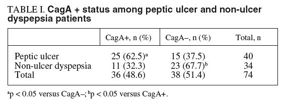
Peptic ulceration (gastric ulcer [n = 5], duodenal ulcer [n = 35]) was detected in 40 patients (54%: 95% CI = 42.1-65.7%, M = 27, F = 13, mean age 40,1 years old, range 18-67), 27.5% being smokers. Thirty-four patients had a normal mucosa or non-erosive endoscopic gastritis (46%, M = 19, F = 15, mean age 41.7% years old, range 20-65), 32% smokers. Thirty six patients (48.6%, 95% CI = 36.8-60.5%, M = 25) were found to be CagA positive on the Elisa assay. A significantly (p < 0.05) higher proportion of peptic ulcer patients had serum CagA positivity: 25/40 (62.5%, 95% CI = 45.8-77.3%), while serum CagA positivity was significantly (p < 0.05) lower among non-ulcer dyspeptic patients: 11/34 (32.3%, 95% CI = 17.4-50.5%)
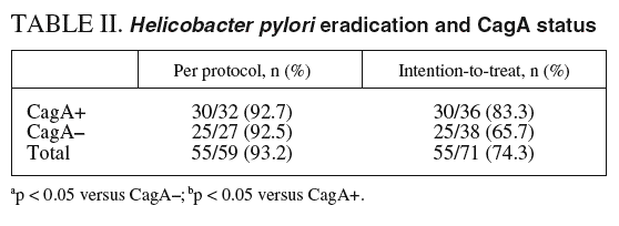
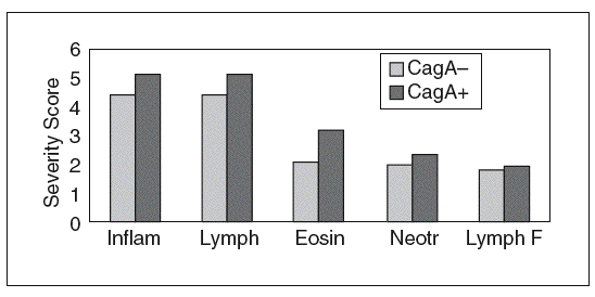
Fifteen patients (peptic ulcer = 10, non-ulcer dyspepsia = 5) refused to be subjected to a repeat endoscopy in order to assess H. pylori eradication. Therefore, eradication was determined in 59 patients (79.7%, 95% CI = 68.8-88.2%). Eradication was successful in 90-95%, on a per protocol analysis, regardless of CagA status (table I), and comparable among peptic ulcer and non-ulcer dyspepsia patients (table II). Histological assessment was performed on samples collected from patients that had H. pylori eradication tested. Therefore, analysis of gastric mucosal inflammation, leukocyte infiltration and development of lymphoid follicules was investigated in 79.7% of patients. The severity of mucosal inflammation was comparable (p = 0.08) among serum CagA positive and negative patients. However, a significantly (p < 0.05) higher eosinophil and lymphocyte mucosal infiltration was observed in CagA positive patients (fig. 1). Similar neutrophil infiltration and lymphoid follicule development was observed among both groups (fig. 1). No differences were observed on the histological severity score when peptic ulcer and non-ulcer dyspeptic CagA positive and negative were analyzed.
Fig. 1. Severity of mucosal inflammation among serum CagA positive and negative patients.
DISCUSSION
A lower eradication rate has been demonstrated among H. pylori positive, non-ulcer dyspeptic patients5-13. Several factors may contribute to this observation, including a reduced severity of gastric mucosal inflammation, likely related to a «less virulent» bacteria. It has been proposed that VacAS2, CagA negative H. pylori infected patients are more resistant to eradication therapy5,10,12,13. Whether this is a result of an increased primary bacterial resistance to antibiotics or to differential drug availability to the gastric mucosa in this particular scenario remains to be elucidated. As consequence, a 10-14 day eradication schedule has been proposed over the commonly employed 7-day triple therapy for peptic ulcer disease, more frequently associated with virulent H. pylori (VacAS1, CagA positive)9.
However, several reports have been published suggesting that H. pylori eradication rates are not influenced by CagA status16-19. Our results showed an eradication efficacy comparable among peptic ulcer and non-ulcer dyspepsia patients, regardless of CagA status. Anycase total numbers are very limited in each group, specially in CagA positive dyspeptic patients, so caution should be taken when interpreting our results. The limited number of patients included can be the explanation for a negative result due to a type B error in the statistic analysis. A high drop-out rate was observed in the study, specially on the CagA negative group. It should be noted that this is a single-center study, and several patients refused to be subjected to a repeat endoscopy.
Gastric mucosal inflammation, characterized by lymphocyte and eosinophil infiltration, was more severe among CagA positive patients. Whether there is a relationship between the degree of mucosal inflammation and the ability of antibiotics to reach the gastric mucosa and the bacteria remains to be elucidated. Therefore, the results of the present study indicate that, despite the presence of H. pylori CagA seropositivity being associated with a more severe gastric mucosal inflammation, H. pylori eradication rates may not be affected by CagA seropositivity status.


