Analizar el valor pronóstico en cuanto a supervivencia de las diferentes características clínicas, histopatológicas e inmunohistoquímicas del carcinoma medular tiroideo (CMT) resecado.
Pacientes y métodoSe han revisado un total de 55 casos de CMT intervenidos consecutivamente y confirmados histológicamente. Los datos referentes a las características clínicas se recogieron de la historia clínica del paciente. Las características histopatológicas e inmunohistoquímicas se obtuvieron del informe anatomopatológico.
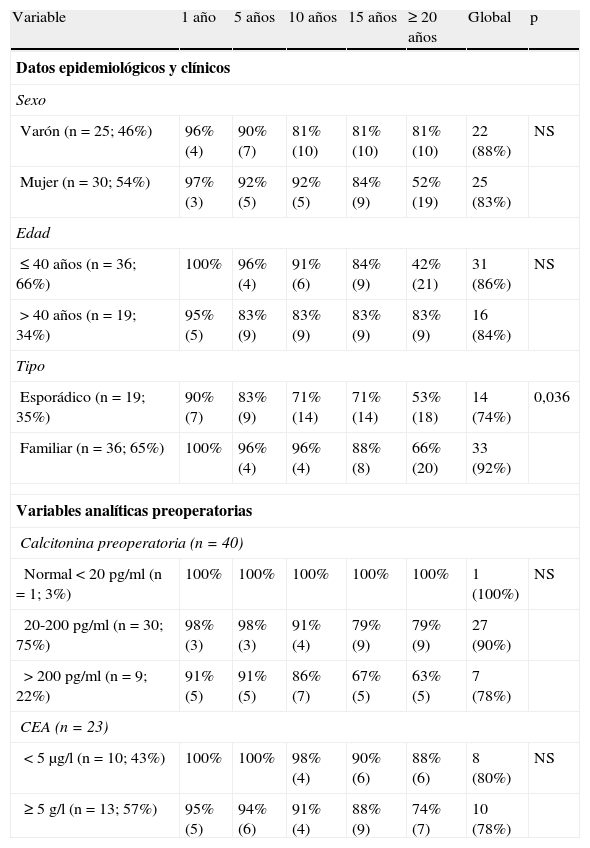
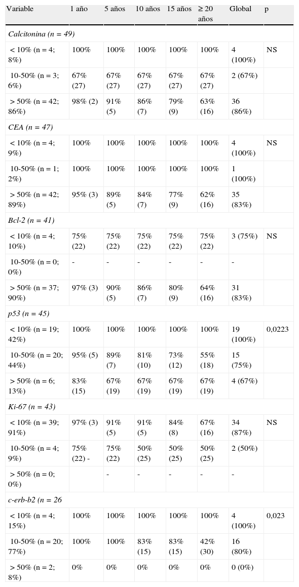
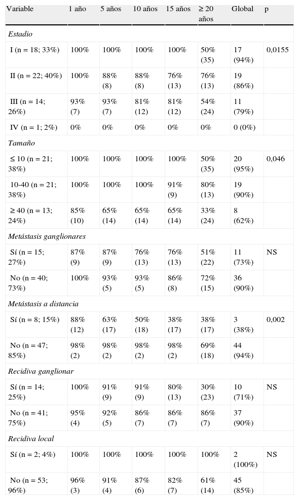
ResultadosLa supervivencia media (DE) al año fue del 96 (2)%; a los 5 años 91 (4)%; a los 10 años 88 (6)%; a los 15 años 83 (7)%, y a los 20 años 61 (14)%. En el análisis de las curvas de supervivencia se obtienen como variables significativas: entre las epidemiológicas, el tipo de tumor (mejor pronóstico el familiar; p=0,035), entre las histopatológicas, la presencia de hiperplasia de células C (p=0,0391) y la presencia de necrosis tumoral (p=0,0005), entre las inmunohistoquímicas, la positividad para p53 (p=0,0223) y para c-erb-b2 (p=0,023), y por último, entre los datos de estadificación, el estadio clínico TNM (p=0,0155), el tamaño (p=0,046) y la presencia de metástasis a distancia (p=0,002). Según el modelo de regresión de Cox, las únicas variables determinantes de mal pronóstico son: la existencia de necrosis (p=0,0391; odds ratio OR 6,5137) y el tamaño tumoral>4cm (p=0,027; OR 14,19659).
ConclusionesLa tasa global de supervivencia del carcinoma medular de tiroides es del 82% y viene determinada principalmente por el tamaño tumoral y por la presencia de necrosis tumoral. Ningún marcador inmunohistoquímico tiene importancia significativa en la supervivencia.
To analyze the importance of various clinical, histopathological and immunohistochemical features in the prognosis of resected medullary thyroid carcinoma.
Patients and methodsA total of 55 cases of medullary thyroid carcinoma consecutively operated were investigated. The data referring to clinical features were collected in the patient's clinical history. The histopathological and immunohistochemical features of the tumors were taken from their pathological anatomy report.
ResultsSurvival at one year was 96±2%; at 5 years 91±4%; at 10 years 88±6%; at 15 years 83±7%; and at 20 years 61±14%. Among epidemiological features, tumor type was significantly related with the disease (best familial prognosis; P=.035); among histopathological features, the presence of C cell hyperplasia and the presence of tumor necrosis had a significant relationship (P=.0005 and P=.039); among immunohistochemical features, positivity for p53 and for c-erb-b2 (P=.023 and P=.022); and finally, among staging data, TNM clinical staging (P=.015), size (P=.046) and the presence of distant metastases (P=.002). According to Cox's regression model, the only variables indicating a poor prognosis were: the existence of necrosis (P=.039; OR=6.513) and tumor size>4cm (P=.027; OR=14.196).
ConclusionsThe survival rate was mainly determined by tumor size and the presence of tumor necrosis. None of the immunohistochemical markers had a significant influence on survival.
Artículo
Comprando el artículo el PDF del mismo podrá ser descargado
Precio 19,34 €
Comprar ahora


