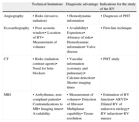
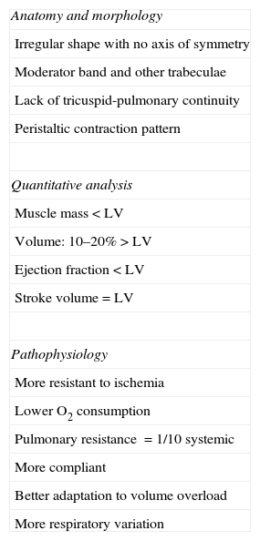
Magnetic resonance (MR) imaging has proven efficacy in the study of the heart. Its clinical applications are directed primarily at the study of the left ventricle, and the right ventricle is relegated to the background. This article reviews the anatomy and physiology of the right ventricle, as well as the manifestations of most common diseases affecting this chamber of the heart: infarction, cardiomyopathy, masses, and right heart failure. Knowing the distinctive features of the right ventricle with respect to the left and the particularities of the MR imaging protocol results in better technical performance in cases in which the reason for the examination or imaging findings point to the right ventricle. The importance of the right ventricle in the management of cardiopulmonary disease is growing and MR imaging can provide clinicians with the support they need.
La resonancia magnética (RM) es una técnica de probada eficacia en el estudio del corazón. Sus aplicaciones clínicas se dirigen preferentemente al estudio del ventrículo izquierdo, quedando el ventrículo derecho relegado a un segundo plano. Este artículo ofrece una revisión de la anatomía y fisiología del ventrículo derecho, así como de las manifestaciones de la afección más frecuente en esta cámara cardíaca: infarto, miocardiopatías, masas y fallo cardíaco derecho. El conocimiento de los rasgos diferenciales del ventrículo derecho con respecto al izquierdo y de las particularidades del protocolo de estudio mediante RM, consigue un mayor rendimiento de la técnica en aquellos casos en que el motivo de petición o los hallazgos de imagen apuntan al ventrículo derecho. La RM reúne características para apoyar desde la imagen el protagonismo creciente que los clínicos están otorgando al ventrículo derecho en el manejo de las enfermedades cardiopulmonares.
Artículo
Comprando el artículo el PDF del mismo podrá ser descargado
Precio 19,34 €
Comprar ahora


