


Información del artículo
Texto completo
Bibliografía
Descargar PDF
Estadísticas
Figuras (8)

Mostrar másMostrar menos
Tablas (5)
Table 1. Cerebral blood flow tests in the process of organ transplant donation.

Table 2. Observational period (whenever the diagnosis of EDa is only clinical).

Table 3. Patients and etiology of the coma (period January 2000–August 2011).
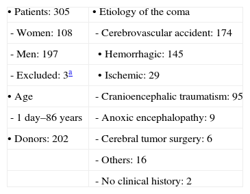
Table 4. Recommendations for performing and interpreting aCPSS.

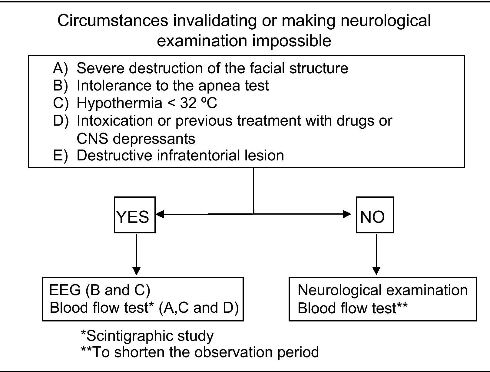
Table 5. Circumstances invalidating or making neurological examination impossible.
Mostrar másMostrar menos
Artículo
Opciones para acceder a los textos completos de la publicación Revista Española de Medicina Nuclear e Imagen Molecular (English Edition)
Suscriptor
Suscribirse

Suscribirse a:
Revista Española de Medicina Nuclear e Imagen Molecular (English Edition)
Comprar
Comprar acceso al artículo
Comprando el artículo el PDF del mismo podrá ser descargado
Precio 19,34 €
Comprar ahora
Comprando el artículo el PDF del mismo podrá ser descargado
Precio 19,34 €
Comprar ahora
Contactar
Teléfono para suscripciones e incidencias
De lunes a viernes de 9h a 18h (GMT+1) excepto los meses de julio y agosto que será de 9 a 15h
Llamadas desde España
932 415 960
Llamadas desde fuera de España
+34 932 415 960
E-mail
