metricas
covid
Buscar en
Revista Española de Medicina Nuclear e Imagen Molecular (English Edition)
Toda la web
Inicio Revista Española de Medicina Nuclear e Imagen Molecular (English Edition) Preliminary evaluation of cerebral 18F-DOPA PET/CT in the differential diagnosis...
Información de la revista
Vol. 40. Núm. 4.
Páginas 214-221 (julio - agosto 2021)
Compartir
Compartir
Descargar PDF
Más opciones de artículo
Visitas
14
Vol. 40. Núm. 4.
Páginas 214-221 (julio - agosto 2021)
Original Article
Preliminary evaluation of cerebral 18F-DOPA PET/CT in the differential diagnosis of brain lesions in inconclusive MR
Valoración preliminar de la 18F-DOPA PET/TC cerebral en el diagnóstico diferencial de lesiones cerebrales con RM no concluyente
Visitas
14
B. Domenech Braseroa,
Autor para correspondencia
beatriz.domenech@quironsalud.es

Corresponding author.
, A. Mestre-Fuscob,c, M. Suárez Piñerac, E. Puertas Calvod, X. Perich Alsinae, G. Montesf, P. Plaza Lópeza
a Servicio de Medicina Nuclear, Hospital Quirónsalud Barcelona, Barcelona, Spain
b Servicio de Medicina Nuclear, Hospital Universitari Doctor Josep Trueta, Girona, Spain
c Servicio de Medicina Nuclear, Hospital del Mar, Barcelona, Spain
d Servicio de Radiodiagnóstico, Hospital Quirónsalud Barcelona, Barcelona, Spain
e Servicio de Oncología Radioterápica, Hospital Quirónsalud Barcelona, Barcelona, Spain
f Departamento de Neurocirugía, Instituto Clavel, Barcelona, Spain
Este artículo ha recibido
Información del artículo
Resumen
Texto completo
Bibliografía
Descargar PDF
Estadísticas
Figuras (2)
Tablas (2)
Table 1. Characteristics of the patients and tumors, reasons for the PET study, previous treatment, current situation and therapeutic attitude.
Table 2. Visual and semiquantitative evaluation; SUVmax, T/S ratio and T/N ratio.
Mostrar másMostrar menos
Abstract
Aim

To evaluate the utility of brain 18F-DOPA PET/CT in the differential diagnosis of brain lesions with inconclusive MRI.
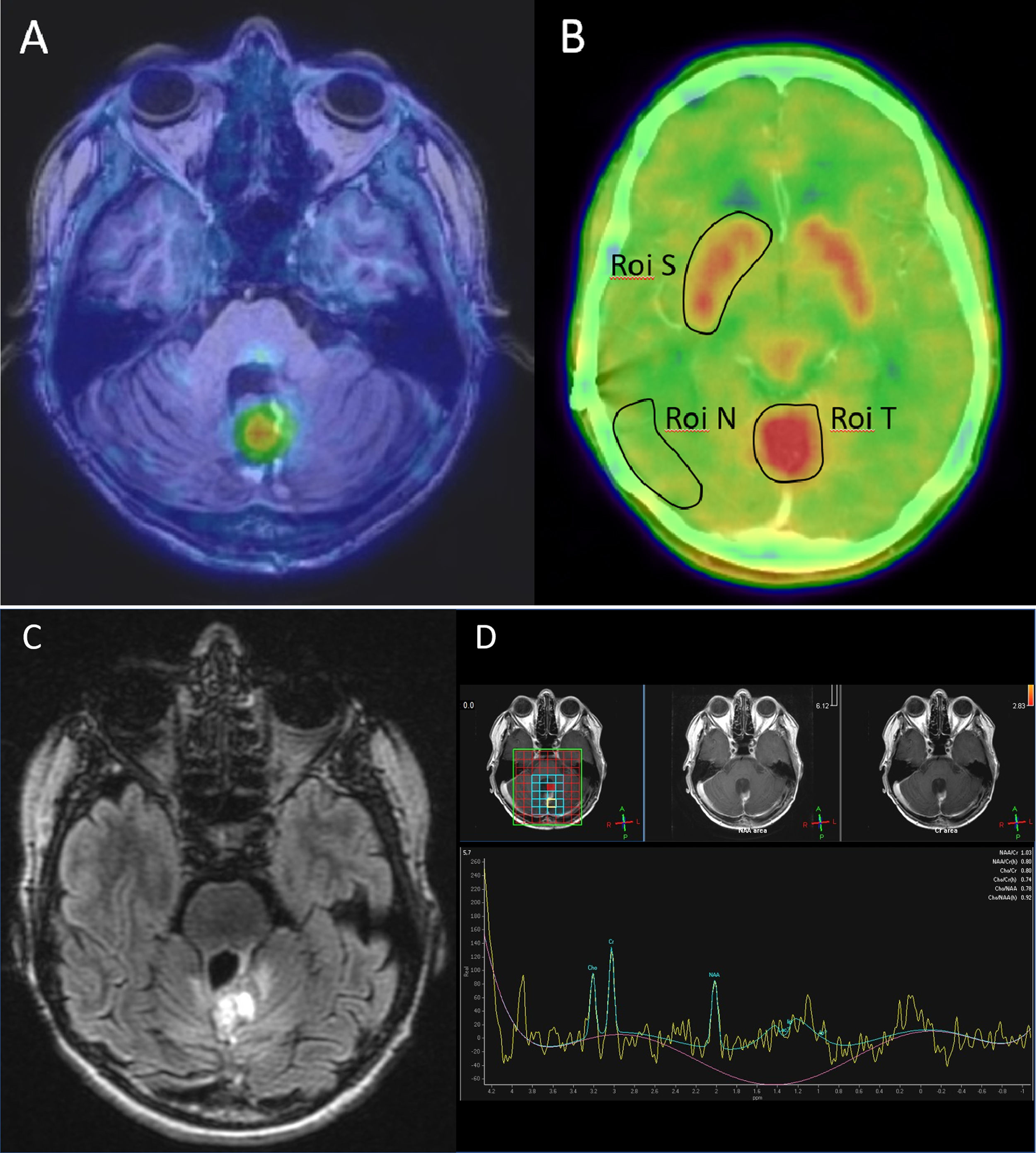
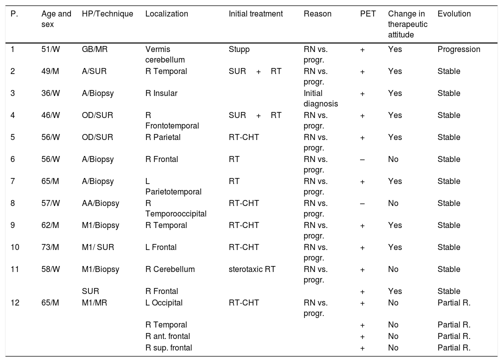
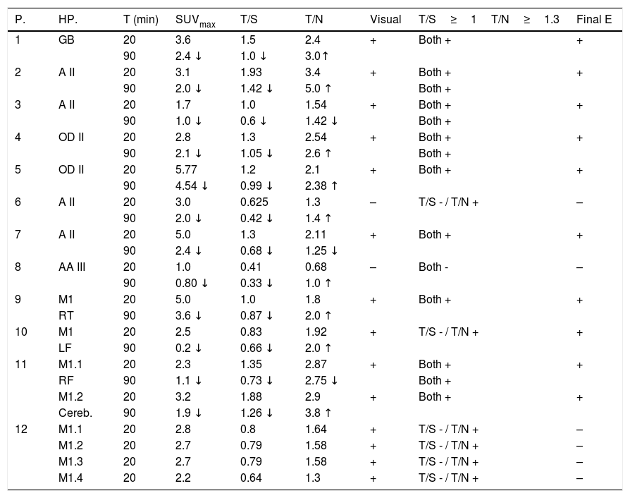
Material and methods

Twelve patients were studied, with a total of 16 lesions, without definitive diagnosis after brain MRI. A double acquisition PET/CT brain scan was acquired at 20 and 90min. Visual and semiquantitative assessment was performed with SUVmax calculation of the lesions and calculation of the T/S Ratio (tumor/contralateral striatum) and T/N Ratio (contralateral healthy tumor/parenchyma) for each time.
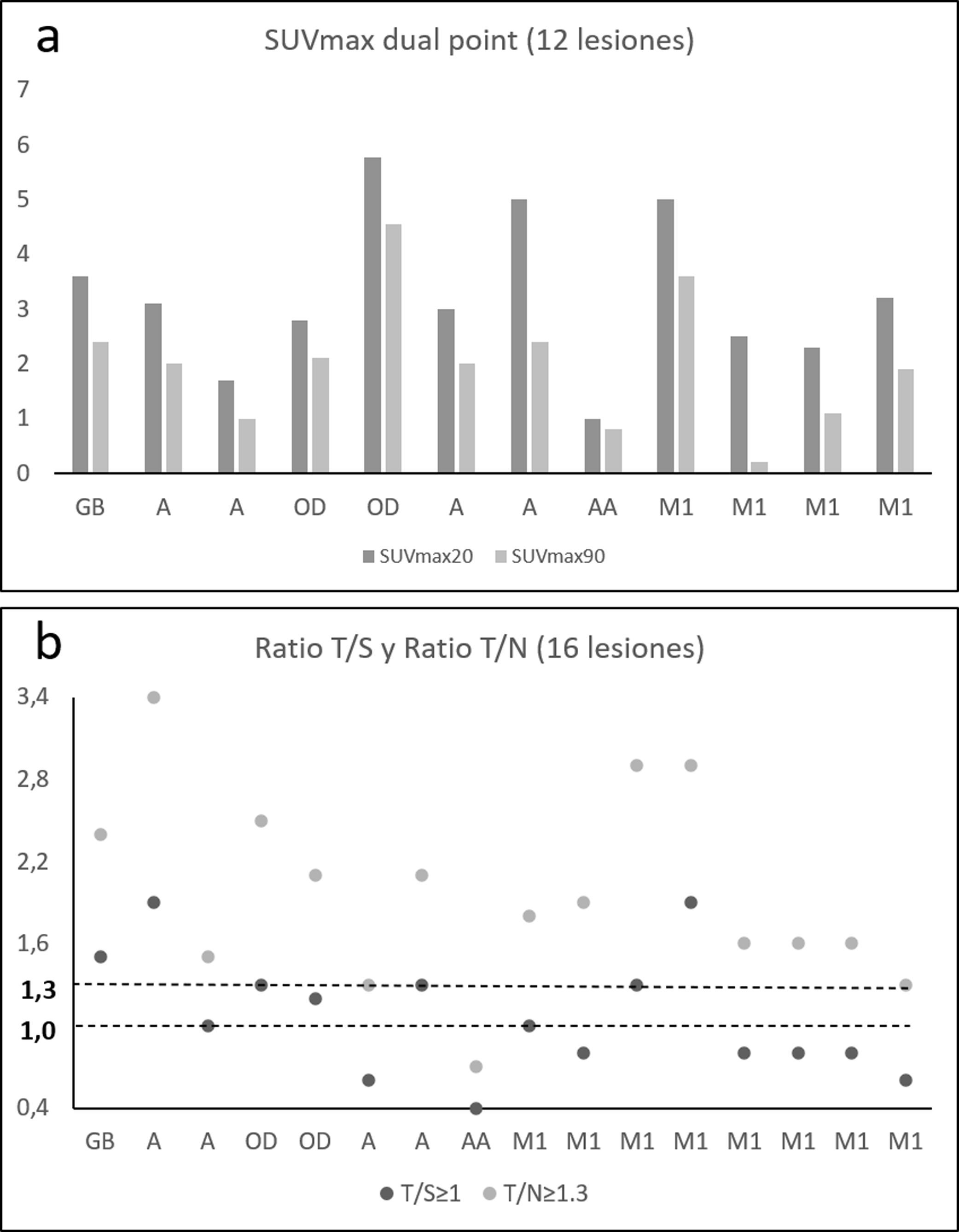
Results

Based on the visual assessment scale and using T/S ratio ≥1 and T/N ratio ≥1.3 to determine malignancy, the values of sensitivity (S), specificity (E) and positive predictive value (PPV) were: visual assessment (S 100%, E 33.3%, VPP 71.4%), T/S Ratio (S 90%, E 100%, VPP 100%) and T/N Ratio (S 100%, E 16.6%, VPP 66.6 %). No lesion showed an increase in SUVmax in late acquisition. 18F-DOPA PET/CT modified treatment in 75% of the patients.

Conclusion

18F-DOPA PET/CT is a useful tool in the study of brain lesions with inconclusive MRI. Late imaging (dual-point) has no added value in the final diagnosis. FDOPA has an impact on patient management modifying therapeutic behavior.

Keywords:
PET/CT
18F-DOPA
Glioma
Brain metastases
Tumoral recurrence
Resumen
Objetivo

Valorar la utilidad de la 18F-DOPA PET/TC cerebral en el diagnóstico diferencial de lesiones cerebrales con RM no concluyente.

Material y métodos

Se estudiaron 12 pacientes, con un total de 16 lesiones, sin diagnóstico definitivo tras RM cerebral. Se adquirió estudio 18F-DOPA PET/TC cerebral con doble adquisición a los 20 y 90minutos. Se realizó valoración visual y semicuantitativa con cálculo del SUVmax de las lesiones y cálculo del Ratio T/S (tumor/estriado contralateral) y Ratio T/N (tumor/parénquima sano contralateral) para cada uno de los tiempos.

Resultados

Basado en la escala de valoración visual y utilizando los ratios T/S1 y T/N1.3 para determinar la positividad de un estudio, los valores de sensibilidad (S), especificidad (E) y valor predictivo positivo (VPP) han sido los siguientes: valoración visual (S 100%, E 33.3%, VPP 71.4%), Ratio T/S (S 90%, E 100%, VPP 100%) y Ratio T/N (S 100 %, E 16.6%, VPP 66.6%). Ninguna lesión mostró un aumento del SUVmax en la adquisición tardía. La 18F- DOPA PET/TC modificó el comportamiento terapéutico en el 75% de los pacientes.

Conclusión

La 18F-DOPA PET/TC es una herramienta útil para diferenciar la etiología de lesiones cerebrales con RM no concluyente. La imagen tardía (dual point) no aporta un valor añadido al diagnóstico final. Los estudios con FDOPA tienen impacto en el manejo del paciente modificando la conducta terapéutica.

Palabras clave:
PET/TC
18F-DOPA
Glioma
Metástasis cerebral
Recidiva tumoral

Artículo

Opciones para acceder a los textos completos de la publicación Revista Española de Medicina Nuclear e Imagen Molecular (English Edition)
Suscriptor
Suscriptor de la revista

Si ya tiene sus datos de acceso, clique aquí.

Si olvidó su clave de acceso puede recuperarla clicando aquí y seleccionando la opción "He olvidado mi contraseña".
Suscribirse
Suscribirse a:

Revista Española de Medicina Nuclear e Imagen Molecular (English Edition)

Comprar
Comprar acceso al artículo

Comprando el artículo el PDF del mismo podrá ser descargado

Precio 19,34 €

Comprar ahora
Contactar
Teléfono para suscripciones e incidencias
De lunes a viernes de 9h a 18h (GMT+1) excepto los meses de julio y agosto que será de 9 a 15h
Llamadas desde España
932 415 960
Llamadas desde fuera de España
+34 932 415 960
E-mail
Opciones de artículo
es en pt

¿Es usted profesional sanitario apto para prescribir o dispensar medicamentos?

Are you a health professional able to prescribe or dispense drugs?

Você é um profissional de saúde habilitado a prescrever ou dispensar medicamentos