La neurolinfomatosis es una manifestación neurológica poco frecuente del linfoma no Hodgkin (LNH), pudiendo ser la primera y única manifestación de la enfermedad. El diagnóstico a menudo es difícil y generalmente requiere la biopsia del nervio, no siendo ésta siempre posible ni concluyente.
Presentamos el caso de una paciente de 66 años, diagnosticada de LNH B de célula grande, en remisión completa por métodos de imagen y biología molecular tras recibir 6 ciclos de quimioterapia. Al cuarto mes tras el tratamiento, la paciente comenzó con un dolor lumbar de características radiculares. Las imágenes TAC y RMN presentaron hallazgos sospechosos de afectación por proceso linfoproliferativo de la raíz L5, objetivando además otras lesiones a nivel torácico.
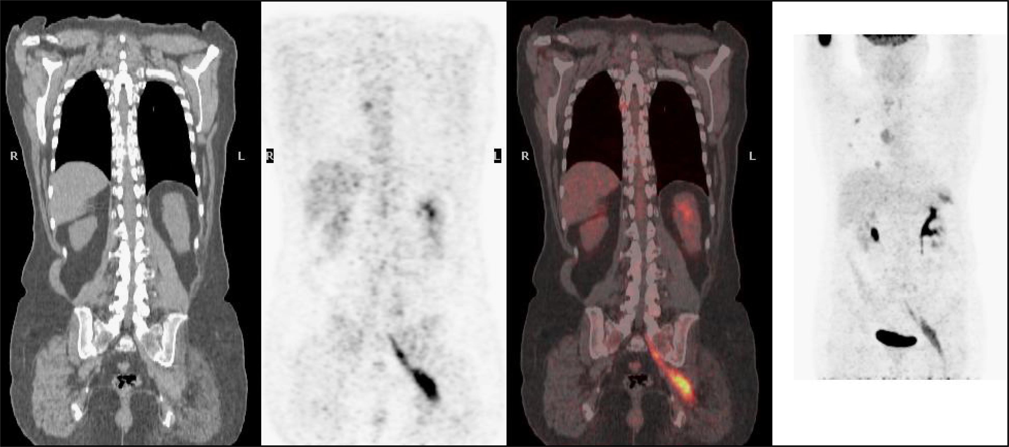
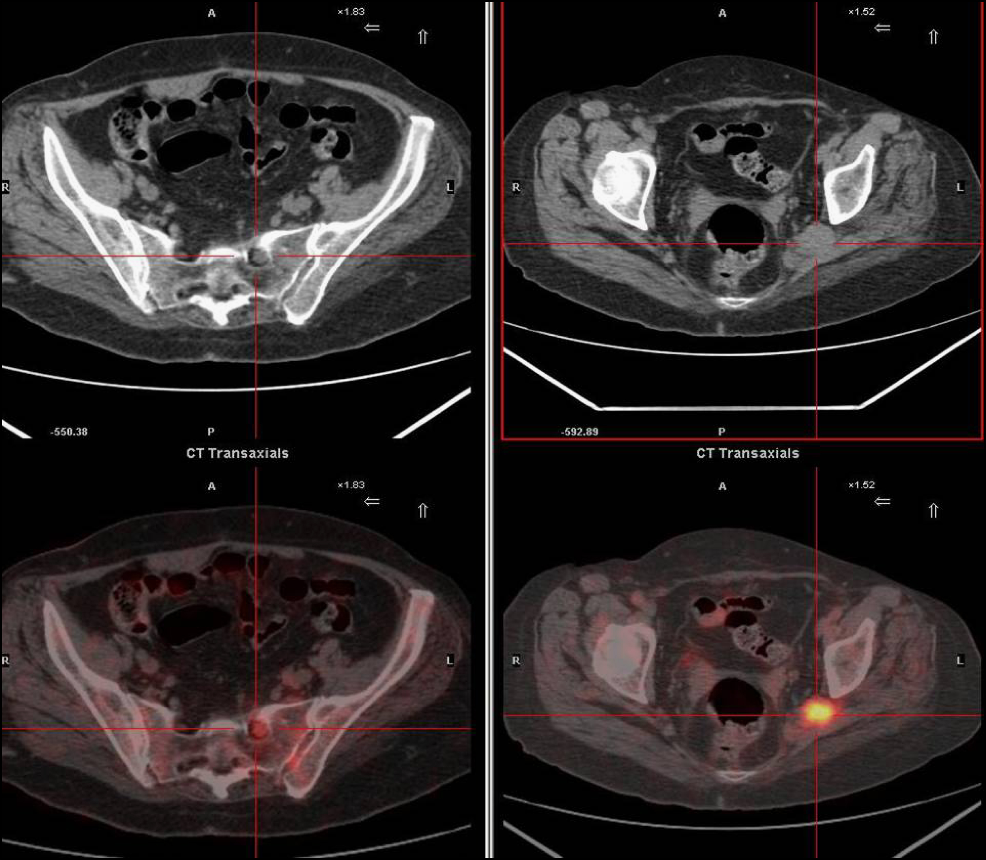
Se solicitó una PET-TAC para completar el estudio y planificar el tratamiento. La exploración confirmó la presencia de recidiva tumoral con infiltración neurolinfomatosa, objetivando además lesiones tanto a nivel torácico como abdominal. Por ello, se decidió iniciar nueva línea de tratamiento quimioterapéutico, prescindiendo de la realización de estudio histológico mediante toma de biopsia.
El caso presentado manifiesta el importante papel que tiene la PET-TAC en el diagnóstico de la neurolinfomatosis. Esta técnica puede evitar la realización de procedimientos agresivos, como el caso del estudio histológico, así como ser de utilidad en el seguimiento y en la valoración de la respuesta al tratamiento de los pacientes diagnosticados de LNH.
Neurolymphomatosis is a rare neurological manifestation of non-Hodgkin's lymphoma (NHL) and it may be its first and sole manifestation. Diagnosis is often difficult and nerve biopsy is generally required. However, this it is not always possible to perform or is not conclusive.
We present the case of a 66-year-old woman diagnosed with giant B-cell NHL. After 6 cycles of chemotherapy, imaging and molecular biology techniques showed complete remission. At four months after treatment, the patient complained of low back pain of radicular distribution. CT and MRI imaging showed signs of lymphoproliferative activity of L5 and also lesions to thoracic nerve roots.
A PET-CT was requested in order to complete the diagnosis and plan the treatment. Imaging confirmed the presence of tumor recurrence with neurolymphomatosis and also indicated lesions on the chest and abdominal level.
Thus, it was decided to start a new line of chemotherapy, without performing the histological study through biopsy.
This case illustrates the important role played by PET-CT imaging in neurolymphomatosis diagnosis. This technique can help the patient avoid more aggressive procedures, such as a biopsy, and can also be useful in the follow-up and assessment of the treatment response to NHL-diagnosed patients.
Artículo
Comprando el artículo el PDF del mismo podrá ser descargado
Precio 19,34 €
Comprar ahora


