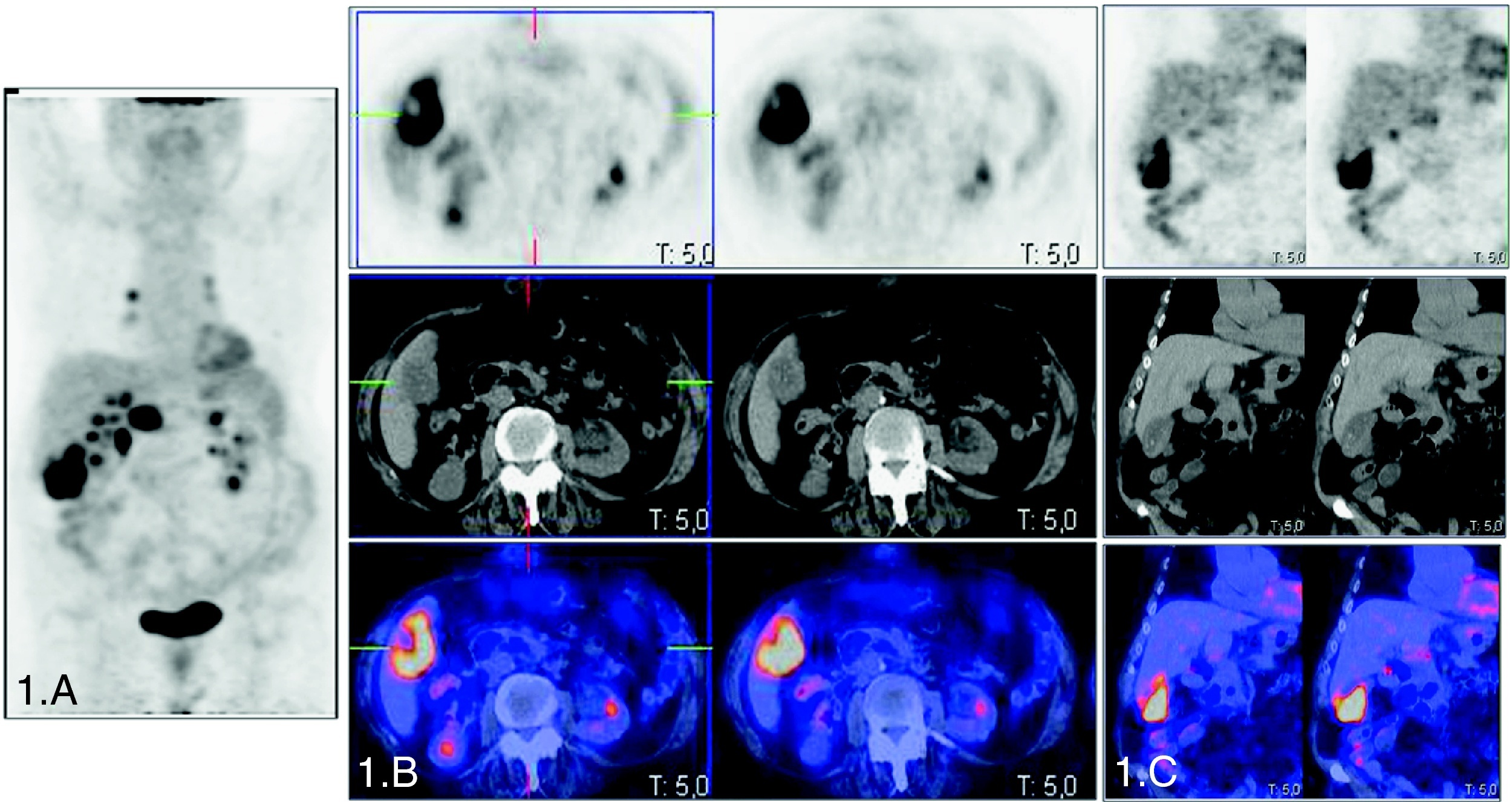
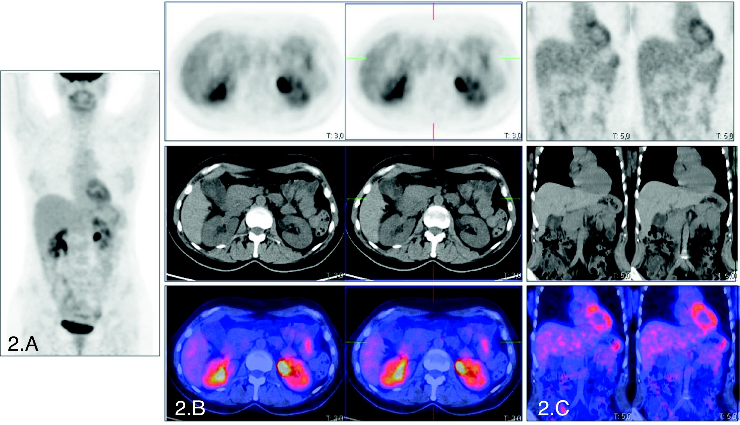
El cáncer de vesícula (CV) es una neoplasia de mal pronóstico en la que el empleo de la tomografía por emisión de positrones (PET) con 18F-fluorodesoxigluosa (FDG) como herramienta diagnóstica puede ser útil, aunque su papel no está bien definido.
Diseño/metodologíaCohorte prospectiva de pacientes con lesión radiológica vesicular sospechosa de malignidad en estudio de diagnóstico y estadificación prequirúrgico mediante PET-FDG en equipos convencionales (PET) y equipos multimodalidad (PET-TAC). Estimación de validez diagnóstica contrastando los resultados de ambos procedimientos con el estudio histopatológico y/o la evolución clínica de los pacientes. Análisis del impacto clínico derivado de su implantación en el diagnóstico del CV.
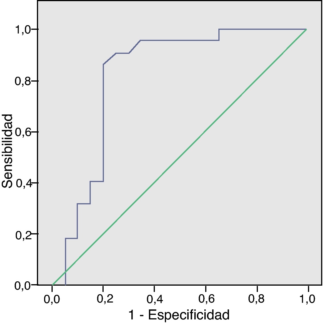
ResultadosSe reclutaron 42 pacientes, 22 con resultado histológico de malignidad y 20 de benignidad. La precisión diagnóstica global fue del 83,33% para el diagnóstico oncológico de la lesión primaria, del 88,89% para la afectación ganglionar y del 85,1% para la afectación metastásica. El SUVmáx medio de las lesiones vesiculares malignas fue 6,14±2,89. La curva ROC mostró un punto de corte de SUVmáx: 3,65 para malignidad. La validez diagnóstica de la PET (n=21) fue discretamente inferior que la de la PET-TAC (n=21). La realización de la PET-FDG modificó la actitud terapéutica en el 14,8%, al encontrar enfermedad diseminada no sospechada.
ComentariosLa PET-FDG diagnostica con precisión la malignidad o benignidad de una lesión vesicular sospechosa, añadiendo la capacidad de identificar enfermedad metastásica no sospechada. La PET-TAC mejora la precisión diagnóstica de la PET por la complementariedad metabólico-estructural de su información. El SUVmáx tiene un valor complementario al análisis visual.
Gallbladder carcinoma is a neoplasm having a poor prognosis in which the role of the positron emission tomography with 18F-fluordeoxyglucose as a diagnostic tool, although of possible usefulness, has not been well-defined.
Methods/designIt is a prospective cohort of patients with radiologically malignant suspicious gallbladder lesions. A staging diagnostic presurgical FDG-PET study was carried out in each patient using both dedicated PET and multimodality PET-CT scanners. Diagnostic accuracy parameters were calculated from the results of PET imaging and were correlated with the condition and/or the clinical course of the patients. The clinical impact of its implementation in the diagnosis of gallbladder carcinoma was also analyzed.
ResultsA total of 42 patients were recruited (22 malignant lesions, 20 benign). Overall diagnostic accuracy was 83.33% for the diagnosis of the primary lesion, 88.89% for the evaluation of lymph node involvement and 85.1% for the evaluation of metastatic disease. Mean SUVmax in malignant gallbladder lesions was 6.14±2.89. ROC curve showed a cut-off value of 3.65 in the SUVmax for malignancy. Accuracy of PET studies alone (n=21) was slightly lower than that of the PET/CT (n=21). FDG-PET changed the management of 14.8% of the population due to the identification of unsuspected metastatic disease.
CommentsFDG-PET accurately diagnoses malignancy or benignity of suspicious gallbladder lesions, with the addition of its capacity to identify unsuspected metastatic disease. PET-CT improves the diagnostic accuracy of the procedure, due to the metabolic-structural complementarity of their information. The SUVmax has a complementary value added to the visual analysis.
Artículo
Comprando el artículo el PDF del mismo podrá ser descargado
Precio 19,34 €
Comprar ahora


