To compare the usefulness of MRI and PET/CT in nodal staging (N) of patients with locally advanced rectal cancer (LARC).
Material and methodsRetrospective study of patients with LARC, who completed their initial staging with PET/CT, between January-20 and March-23. Regional nodes were assessed, and N was determined using both techniques according to TNM criteria. Concordance between MRI and PET/CT was analyzed. The accuracy of both techniques was calculated for those patients who underwent direct surgery. Non-regional pelvic lymph nodes were evaluated by both modalities.
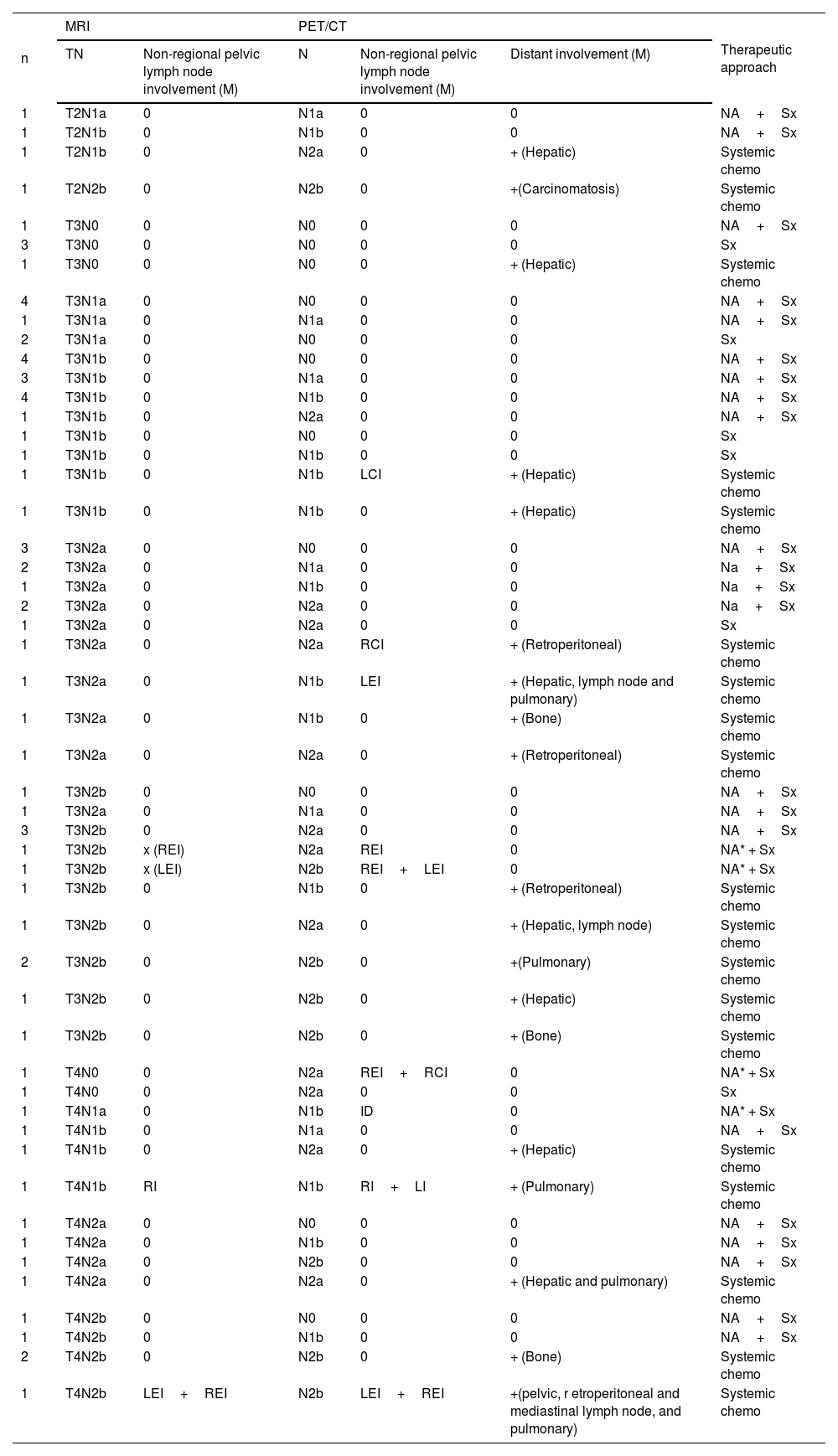
ResultsAmong the 73 patients, 48 were ultimately diagnosed with a locally advanced stage. Of these, 39 underwent neoadjuvant treatment (chemoradiotherapy) followed by surgery, and 9 direct surgery. In 25, the PET/CT extension study revealed distant disease, leading to systemic treatment. Weak concordance was observed between MRI and PET/CT in determining N (k=0.286; p<0.005). Out of 73 patients, 31(42%) exhibited concordance, and 42(58%) showed discordance. In 83% of the discordant cases, MRI overstaged compared to PET/CT, with 17 cases indicating nodal involvement (N+) by MRI and N0 by PET/CT. Diagnostic accuracy was 78% for both techniques. Sensitivity, specificity, positive predictive value, and negative predictive value were 80%, 75%, 80%, and 75% for MRI, and 60%, 100%, 100%, and 67%, for PET/CT. PET/CT identified pelvic metastatic adenopathies in 8 patients that were not visible/doubtful by MRI.
ConclusionsIn the initial nodal staging of rectal cancer MRI overstages relative to PET/CT. Both modalities are complementary, PET/CT offers higher specificity and MRI higher sensitivity.
Comparar la utilidad de la RM y la PET/TC en la estadificación ganglionar (N) de pacientes con cáncer de recto localmente avanzado (CRLA).
Material y métodosEstudio retrospectivo de pacientes con CRLA, que completaron su estadificación inicial con PET/TC, entre enero-20 y marzo-23. Se valoraron los ganglios locorregionales, determinando N por ambas técnicas según TNM. Se analizó la concordancia entre RM y PET/TC. Se calculó la exactitud diagnóstica(ED) de ambas técnicas en aquellos pacientes sometidos a cirugía directa. Se evaluó la afectación ganglionar pélvica no locorregional por ambas modalidades.
ResultadosDe 73 pacientes, 48 presentaron finalmente un estadio localmente avanzado, de éstos, 39 fueron tratados con neoadyuvancia (Quimio-radioterapia) y cirugía, y 9 con cirugía directa. En 25 pacientes al completar el estudio de extensión mediante PET/TC se encontró enfermedad a distancia, por lo que recibieron tratamiento sistémico. Se obtuvo concordancia débil entre RM y PET/TC para determinar N (k=0,286; p<0,005), con 31/73 (42%) concordantes y 42/73 (58%) discordantes. De los discordantes, en 35/42 (83%) la RM supraestadificó en relación al PET/TC, siendo 17 de estos N+(RM) y N0 (PET/CT). Valores de ED: 78% para ambas técnicas. S, E, VPP y VPN: del 80%, 75%, 80% y 75% para RM, y 60%, 100%, 100% y 67% para PET/CT. En 8 pacientes PET/TC identificó adenopatías metastásicas pélvicas no visibles/dudosas por RM.
ConclusionesEn la estadificación ganglionar inicial del cáncer de recto la RM supraestadifica en relación al PET/TC. Ambas modalidades son complementarias, PET/TC ofrece mayor especificidad y RM mayor sensibilidad.
Artículo

Revista Española de Medicina Nuclear e Imagen Molecular (English Edition)
Comprando el artículo el PDF del mismo podrá ser descargado
Precio 19,34 €
Comprar ahora


