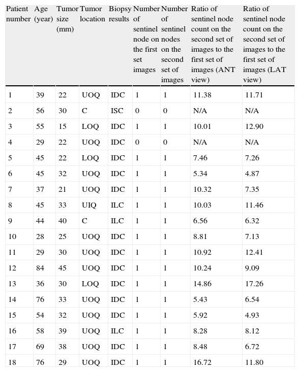
A major controversial issue in the sentinel node biopsy of the breast is the applicability of sentinel node mapping in patients with the history of previous excisional biopsy of the breast lesions. In the current study, we evaluated the reproducibility of lymphoscintigraphy before and after excisional biopsy of the primary breast lesions using superficial peri-areolar injection of the radiotracer.
Material and methodsEighteen patients scheduled for excisional biopsy of breast lesions were included into the study. The patients received intra-dermal injection of the radiotracer in the peri-areolar area of the index quadrant 1 to 2h before surgery. Imaging was performed the day after surgery. Immediately after completion of the first imaging, the patients received another injection of the radiotracer with the same technique, dose, and location. Other sets of lymphoscintigraphy imaging were taken immediately and 4h post second injection. The two sets of lymphoscintigraphy images were compared.
ResultsIn 2 patients, sentinel node could not be identified in either set of images. In the remaining 16 patients, one sentinel node was detected in both lymphoscintigraphy image sets. The sentinel nodes of the second image sets were all in the same location of the first sets with at least 5 times higher count.
ConclusionsExcisional biopsy of the primary breast lesions does not seem to change the superficial lymphatic drainage pattern from the areola of the breast and sentinel node mapping can be performed after this procedure using superficial periareolar technique.
Una cuestión de gran controversia en la biopsia del ganglio centinela de la mama es la aplicabilidad del estudio del ganglio centinela en pacientes con historia previa de biopsia excisional de las lesiones de la mama. En el presente estudio, evaluamos la reproducibilidad de la linfogammagrafía antes y después de la biopsia excisional de las lesiones primarias de mama utilizando la inyección periareolar superficial del radiotrazador.
Material y métodosSe incluyó en el estudio a 18 pacientes programadas para biopsia excisional de lesiones de mama. A las pacientes se les administró una inyección intradérmica del radiotrazador en el área periareolar del cuadrante con tumor, con 1 o 2h antes de la cirugía. La imagen se obtuvo el día posterior a la operación. Inmediatamente tras la primera imagen, a las pacientes se les administró otra inyección del radiotrazador con la misma técnica, dosis y localización. Se realizaron inmediatamente otras series de imágenes de linfogammagrafía, y a las 4h después de la segunda inyección. Se compararon las 2 series de imágenes de linfogammagrafía.
ResultadosEn 2 pacientes no se pudo identificar el ganglio centinela en ninguna de las series de imágenes. En las 16 pacientes restantes se detectó un ganglio centinela en ambas series de imágenes de linfogammagrafía. Los ganglios centinela de las segundas series de imágenes se detectaron en la misma localización que las primeras series de imágenes, con un contaje al menos 5 veces superior.
ConclusionesLa biopsia excisional de las lesiones primarias de mama no parece modificar el patrón del drenaje linfático superficial desde la areola de la mama, pudiendo realizarse el estudio del ganglio centinela tras esta intervención, utilizando la técnica periareolar superficial.
Artículo

Revista Española de Medicina Nuclear e Imagen Molecular (English Edition)
Comprando el artículo el PDF del mismo podrá ser descargado
Precio 19,34 €
Comprar ahora


