The aim of this study was to evaluate whether tumor standardized uptake value (SUVmax) and metabolic tumor volume (MTV) associate with the presence of PET-positive pelvic/para-aortic lymph nodes (LN) in cervical cancer patients.
MethodSeventy-four patients with stage IB-IVB cervical cancer (squamous [n:66], nonsquamous [n:8]), who were referred to FDG-PET/CT department for initial staging, were enrolled in this study.
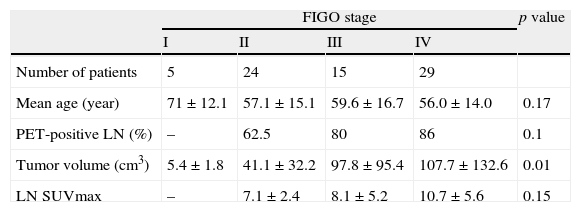
ResultsPatients were staged according to International Federation of Gynecology and Obstetrics [FIGO] criteria as; stage I (n:5), stage II (n:25), stage III (n:15) and stage IV (n:29). PET/CT detected 53 patients with hypermetabolic LN (average SUVmax: 7.5±4.1, range: 4.1–22.8, pelvic LN: 29 patients, para-aortic LN:5 patients, pelvic and para-aortic LN:19 patients). SUVmax and MTV were significantly higher in patients with PET-positive LN compared to others (18.4 and 88.8cm3 vs. 13.9 and 39.9cm3 respectively, p=0.007 for SUVmax, p=0.0001 for MTV). Cut-off values in association with PET-positive LN were 15.2 for SUVmax and 35cm3 for MTV on ROC curve analysis. There was no correlation between SUVmax and MTV (correlation coefficient (R2)=0.07). MTV differed significantly with FIGO stages (41, 98 and 107cm3, in stage II, III and IV respectively, p=0.015).
ConclusionPresence of PET-positive LN correlates with tumor SUVmax and MTV of cervical tumor. These findings support the use of PET/CT in the pretreatment evaluation of cervical cancer patients in order to identify cases with high risk of lymphatic involvement.
El objetivo de este estudio fue el de evaluar si el valor de captación tumoral estándar (SUVmax) y el volumen tumoral metabólico (VTM) se asocian a la presencia de ganglios linfáticos pélvicos/para-aórticos PET-positivos en los pacientes de cáncer cervical.
MétodoSe incluyó en este estudio a setenta y cuatro pacientes con cáncer cervical en estadio IB-IVB (escamoso [n:66], no escamoso [n:8]), que fueron remitidos a la unidad de FDG-PET/TC para una estadificación inicial.
ResultadosSe clasificó a los pacientes de acuerdo a los criterios de la Federación Internacional de Ginecología y Obstetricia [FIGO], es decir; estadio I (n:5), estadio II (n:25), estadio III (n:15) y estadio IV (n:29). La exploración PET/CT detectó 53 pacientes con ganglios hipermetabólicos (SUVmax medio: 7,5±4,1, rango: 4,1-22,8, ganglios pélvicos: 29 pacientes, ganglios para-aórticos: 5 pacientes, ganglios pélvicos y para-aórticos: 19 pacientes). Los valores de SUVmax y VTM fueron considerablemente superiores en pacientes con ganglios PET-positivos en comparación al resto (18,4 y 88,8cm3 frente a 13,9 y 39,9cm3 respectivamente, p=0,007 para SUVmax, p=0,0001 para VTM). Los valores límite en cuanto a ganglios PET-positivos fueron de 15,2 para SUVmax y 35cm3 para VTM en un análisis de curva ROC. No se produjo correlación entre SUVmax y MTV (coeficiente de correlación: (R2)=0.07). El VTM difirió significativamente de los estadios FIGO (41, 98 y 107cm3, en los estadios II, III y IV respectivamente, p=0,015).
ConclusiónLa presencia de ganglios PET-positivos guarda correlación con el SUVmax tumoral y el VTM del tumor cervical. Estos hallazgos apoyan el uso de PET/TC en la evaluación previa al tratamiento de los pacientes de cáncer cervical, a fin de identificar los casos con elevado riesgo de implicación linfática.
Artículo

Revista Española de Medicina Nuclear e Imagen Molecular (English Edition)
Comprando el artículo el PDF del mismo podrá ser descargado
Precio 19,34 €
Comprar ahora


