This study has aimed to evaluate the impact of F18 Fluorodeoxyglucose Positron Emission Tomography-Computed Tomography (FDG PET-CT) in the differential diagnosis of malignant and benign pleural lesions in patients with suspected malignant pleural mesothelioma (MPM).
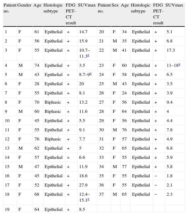
Material and methodsFifty patients (32 females, 18 males; age range 24–79 years) with pleural thickening, fluid, plaques or calcification on previous CT scan were examined with FDG PET-CT. PET-CT imaging was obtained 1h after FDG injection. In 12 patients, delayed imaging from the thoracic region was performed 2h after injection. FDG uptake was evaluated visually and semiquantitively using standardized uptake value (SUV). FDG PET-CT findings were compared with histopathologic diagnosis.
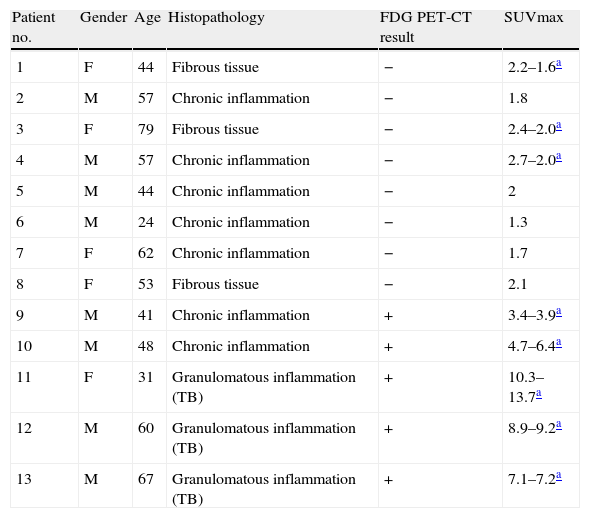
ResultsThirty-nine patients had increased FDG uptake in pleural lesions but PET-CT results were negative in 11 patients. When compared with histopathological results in FDG positive group, 34 patients had MPM, 5 had benign pathology; in FDG negative group 8 patients had benign pathology, 3 had MPM. Of patients with delayed imaging, 9 showed increased SUV but 3 had a decreased SUV on delayed images. Increased SUV group had 4 MPM, 5 benign pathology (3 chronic granulomatous inflammation, 2 benign asbestotic plaque). Decreased SUV group all had benign pathology (fibrosis, chronic inflammation, myofibrosis).
DiscussionFDG PET-CT is a useful imaging modality in differential diagnosis of malignant and benign pleural lesions. Delayed imaging seems to be useful if there is a decrease in SUV suggesting a benign pathology but does not seem to contribute to the differential diagnosis if the SUV is increased.
El propósito de este estudio fue el de estudiar el impacto de F-18 Fluoro Desoxi Tomografía-Tomografía por Emisión de tomografía computerizada (FDG y PET-TAC) sobre la diagnosis diferencial de lesiones benignas y malignas en pacientes con sospecha de mesotelioma pleural maligno.
Pacientes y métodosCincuenta pacientes (32 mujeres, 18 hombres, rango de edad 24-79 años, edad media 57,6) con engrosamiento pleural, derrame pleural, placas o calcificaciones vistas en imágenes en estudios previos con TAC fueron sometidos a la PET-TAC con FDG. Tras la inyección intravenosa de la FDG, se realizaron imágenes PET-TAC al cabo de una hora. Se obtuvieron imágenes tardías de la región torácica a las 2 horas de la inyección. Se llevaron a cabo análisis visual y semi cuantitativos con el valor de captación estándar (SUV). Los resultados del FDG PET-TAC fueron comparados con el diagnóstico histopatológico.
Resultados39 pacientes demostraron captación aumentada en lesiones pleurales. Los resultados en la PET-TAC fueron negativos en 11 pacientes. Al comparar los resultados histopatológicos en el grupo de FDG-positivo, 34 pacientes tenían mesotelioma pleural maligno (MPM), 5 pacientes tenían patologías benignas. En el grupo FDG-negativo, las patologías fueron benignas en 8 pacientes, 3 pacientes tuvieron MPM. En los pacientes con imágenes tardías, 9 demostraron SUV aumentado, pero 3 tuvieron SUV disminuido en las imágenes tardías. En el grupo con SUV aumentado, 4 tuvieron MPM, 5 patología benigna (3, inflamación granulomatosa, y 2 placas asbestosis benignos). En el grupo de SUV disminuido, todos tuvieron patología benigna (la fibrosis, inflamación crónica, miofibrosis).
DıscusıónLa FDG PET-TAC es una modalidad de imágenes útil en el diagnóstico diferencial de lesiones pleurales benignas y malignas. Imágenes tardías parecen ser útiles si existe una disminución en SUV sugerente de una patología benigna. Sin embargo, no parece contribuir a un diagnóstico diferencial si el SUV se aumenta.
Artículo

Revista Española de Medicina Nuclear e Imagen Molecular (English Edition)
Comprando el artículo el PDF del mismo podrá ser descargado
Precio 19,34 €
Comprar ahora


