


The purpose of this study was to evaluate the displacement of the condyle and dental arches with the record of an immediate deprogramming splint one month after the use of the splint in TMJ symptomatic patients.
Material and methodsThe records of 15 symptomatic patients who were subjected to a questionnaire and clinical evaluation for their inclusion in the study were used. Casts were mounted from three records: 1) maximum intercuspation, 2) centric relation with Roth power centric bite, and 3) centric relation a month after the daily use of the splint. Condylar position of the right and left side in horizontal and vertical was measured and compared as well as the vertical and horizontal overbite in each of the three records.
ResultsThere were no significant differences in the results obtained from the records made in centric relation with an immediate splint and in centric relation after the use of the occlusal splint. However, it was found that there is a considerable discrepancy in the condylar and dental displacement in a position of maximum intercuspation and in centric relation after the use of the splint, as well as an improvement in the patients’ symptoms with the use of that device.
ConclusionsCast mounting on the articulator is recommended for diagnostic purposes, in the measurement of condylar position and the use of splint in patients with TMJ pain to identify more reliably the goals to consider during and by means of orthodontic treatment. Otherwise they could be masked and determine a different course in treatment planning.
El propósito de este estudio fue el de evaluar el desplazamiento condilar y de las arcadas dentales con el registro de un desprogramador inmediato, al mes posterior del uso de una guarda oclusal en pacientes sintomáticos de ATM.
Material y métodosSe utilizaron los registros de 15 pacientes sintomáticos que fueron sometidos a un cuestionario y evaluación clínica para su inclusión. Se montaron los modelos a partir de tres registros: 1) máxima intercuspidación, 2) relación céntrica con power centric bite de Roth, y 3) relación céntrica un mes posterior al uso diario de una guarda oclusal. Se midió y comparó la posición condilar de los lados derecho e izquierdo en sentidos horizontal y vertical, así como la sobremordida vertical y horizontal en cada uno de los tres registros.
ResultadosNo existen diferencias significativas en los resultados obtenidos a partir de los registros realizados en relación céntrica con un desprogramador inmediato y relación céntrica posterior al uso de la guarda oclusal. Sin embargo, se comprobó que existe una discrepancia considerable en el desplazamiento condilar y dental en una posición de máxima intercuspidación a relación céntrica posterior al uso de la guarda, así como una disminución de la sintomatología en pacientes con el uso de dicho dispositivo.
ConclusionesSe recomienda el uso del montaje de modelos en articulador con fines de diagnóstico, en la medición de la posición condilar y la utilización de guarda en pacientes con dolor de ATM, para identificar de manera más confiable los objetivos a considerar durante y por medio del tratamiento ortodóncico, de otra manera éstos pudiesen encontrarse enmascarados y determinarían un curso distinto en la planeación del mismo.
In regard to any dental procedure, the mandible may assume two positions as reference for executing a treatment plan: centric relation (CR) and maximum intercuspation (MI).1
CR is considered the most reliable and reproducible point of reference to register the precise relationship of the mandible with the maxilla,1 where the joints can withstand loads without causing discomfort or unease. On the other hand, MI is defined as the occlusal relation in which the teeth of both arches fully interpose.2 Several authors and -professors and clinicians- recognized by their understanding of gnathologic function, coincide in the assertion of considering CR as a physiological goal in orthodontic correction. Therefore, determining this position is a prerequisite for interarch dental analysis, condyle position and skeletal relationship.1
Cordray states that CR does not usually coincide with the mandibular position obtained in MI. Thus there is a difference between the three-dimensional position of the condyle in the CR (condylar position when they are seated) and MI (dictated by the occlusion). This phenomenon was given the name of condylar displacement.1 The displacement direction depends on how the occlusion is altered.3 This significant change in interarch dental position can affect diagnosis and orthodontic treatment since the magnitude of the dental and skeletal discrepancy, probably caused by the same problem, is much more evident. Therefore, a comprehensive assessment of the occlusion must include the study of cast models mounted in CR, and also of the condylar position resulting from dental maximum intercuspation.1 An objective method for measuring condylar position is the use of instruments designed for this purpose such as the CPI (condylar position indicator). This tool has apparently proved to be accurate and reliable.4
In order to properly seat the condyles and study the dental arches, as well as changes in their positions, a method that reduces or eliminates the influence of occlusion over the musculature must be used. By doing so, dental interference might be sidestepped to achieve correct positioning of the mandible and closure without considering condylar position.1 The constant repetition of signals to proprioceptive receptors in the muscles causes the establishment of a deviation pattern called engram.5Therefore, clinical handling is unreliable to determine CR. Calagna et al., determined that neuromuscular deprogramming could be the key to achieving reproducibility of mandibular position,6 while Cordray1 states that the clinician cannot assume that the condyles are seated before treatment only because the patient is asymptomatic.1
Under these assumptions, several methods have been developed to achieve the immediate deprogramming among which is the Roth modified power centric bite record. This record uses the muscles that provide closure to seat the condyles closest to the CR.7
However, to study the influence of condylar position dictated by occlusion in symptomatic patients it is necessary to use a method that efficiently reduces or eliminates the influence of occlusion over the neuromusculature.4 The therapy with occlusal splints recommended by Roth may be effective for such deprogramming and it may be the ideal protocol for achieving a reliable CR record.2,3 This therapy consists in the construction of an individualized splint with features such as immediate canine disocclusion and mutually protected occlusion. The splint is adjusted as required until changes in mandibular position are no longer registered. Additionally, it is an element for diagnosis and an adjunctive therapy for pain relief of TMJ symptomatic patients.4
Tamburrino et al.3 conducted a study to investigate the magnitude of dental displacement and occlusal changes related to the magnitude of the corresponding condylar displacement. However, the study was carried out in patients 7 to 17 years without the use of an occlusal splint -even in cases of difficult handling and with symptomatology- to obtain a record of centric relation.
To date, no research has been conducted that compares the obtained results with the Roth’s5 CR registration technique and CR after the use of an occlusal splint.
Material and methodsThe study reviewed 30 patients, from which a sample of 15 was obtained, of both genders, with an age between 16 and 55 years, with TMJ symptoms; dental Angle Class I, without prior orthodontic treatment; with complete permanent dentition including second molars; non-carriers of prostheses, without medical conditions that would prevent their inclusion in the study and without psychomotor limitations. The patients were not in treatment with antidepressants and had no third molars present.
To find out if the patients were symptomatic a questionnaire based on the evaluation of Helkimo was used to categorize and quantify the subjectivity of the TMJ symptoms. We included patients who had a score above the unit according to the scale of Helkimo.
The statistical technique of Helkimo consists in quantifying the results obtained from prior examinations and clinical anamnesis in relation to the severity degree. The patients personally answered a questionnaire that assessed the presence of symptoms in various areas of head/neck.
Clinical examMedical and dental care questionnaires were performed as well as the clinical evaluation based on the criteria established by Helkimo. All subjects were examined in order to confirm the presence of conditions in mandibular movement and joint function, masticatory muscle pain, articulation and attrition.
To evaluate mandibular function and joint pain each subject was asked to open their mouths as much as possible. Maximum opening was measured and deviations or pain were registered. Maximum opening was defined as the distance between the upper and lower incisal edges minus the amount of overbite. The patients were asked to perform lateral movements, recording any pain in the joint or in the muscles. Then, the following muscles were palpated bilaterally: temporalis (anterior, posterior and middle); masseter and internal pterygoid superficial and deep fascia. To reduce the possibility of error muscles that could easily be isolated were included. For this reason, the lateral pterygoid was not included. Finally, the joints were palpated through the external auditory meatus during closing and opening.
After performing the assessment, a sample of 15 individuals that showed some sign or symptom was selected.
The following procedure was performed with each of the individuals:
- 1.
Perforated impression trays (TP Orthodontics) and alginate were used (Orthoprint, Zhermack, Italy) for all the impressions. All models were obtained with type IV plaster immediately after taking the impressions (Vel-Mix, Kerr Manufacturing Co., Romulus, Mich).
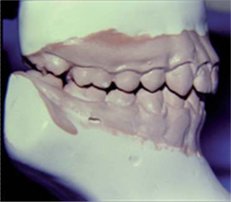
The models were mounted using the split cast technique in a Panadent articulator (Panadent Corp, Grand terrace, CA) following the procedure recommended by the manufacturer. The upper models were mounted with the aid of a facial arch manufactured by the same company using mounting plaster (Figure 1).
- 2.
We measured the condyle’s position in the horizontal (X) and vertical (Y) axis using the CPI (Model CPI, Panadent Corp, Grand terrace, CA). To reduce the effect of errors in the measurement, three separate measures were carried out for each subject and the results were averaged. All measurements were taken with a 10x magnifying glass (Figure 2).
 Figure 2.
CPI record.
Overjet. Distance measured in milimeters from the buccal surface of the lower central incisor to the lingual surface of the upper central incisor, tangent to the incisal edge of the latter.
Overbite. Distance measured in milimeters from the incisal edge of the upper central incisor to the incisal edge of the lower central incisor.
(0.06MB). - 3.
For dental analysis the models were examined with a 300mm/12 Digitaces (Kai Shi International Co., Ltd) electronic caliper.
- 4.
A bite record in CR was obtained through immediate deprogramming with Roth’s modified power centric bite. A new pair of models was mounted and the same skeletal and dental aspects mentioned above were analyzed.


- 1.
The patient -positioned in the dental chair at a 45 degree angle from the floor- was instructed to bite continuously (every 5 seconds) a wooden tongue depressor with a moderate and pulsatile force for about 5-10 min to temporarily modify the engram.
- 2.
The wax’s anterior portion (4 widths) was placed on the anterior teeth and was held with the index fingers over the buccal surfaces. Using the thumb, the operator guided the mandible softly (not to manipulate but to avoid mandibular protrusion); the patient was told to close slowly with the posterior teeth until a 2mm disoclussion was obtained between the upper and lower posterior teeth. The patient was asked to hold that position. The wax was cooled down with air until it hardened in order to avoid deformations upon removal. The wax was then placed in a glass with cold water. The patient was not allowed to close until all records were taken.
- 3.
The posterior portion of the wax (2 widths) was placed on the upper teeth and was held with the index fingers over the buccal surfaces. With the posterior portion of the wax inside the mouth, the anterior wax was placed again with no contact of the posterior teeth.
- 4.
With the free hand, the mandible was guided to CR. The lower anterior teeth should then reach the previous indentation without any shift. The patient was asked to close and remain in that position. The condyles seated as they closed on the anterior stop and the posterior dental axis closed without resistance. The posterior wax was cooled down with air and both waxes were removed upon complete hardening and placed in cold water.
- 5.
The occlusal splint was built with anterior guidance, with no posterior contacts during protrusion or lateral movements and with equal bilateral multiple contacts in CR. Continuous wear during 30 days was prescribed. Weekly adjustments were made by removing premature contact points or adding acrylic depending on each case.
The statistic test used in this study was the nonparametric H-test or Kruskal-Wallis test which allowed the comparison of two independent experimental groups at different times to determine if there were statistical significant differences between them.
This method is adequate to compare populations with abnormal distributions and when the sample’s standard deviations differ considerably. In the H test the sample observationsarereplaced withtheircombined ranges, so that range 1 is assigned to the smallest of the combined observations and the average of the ranges is assigned to tie values that otherwise would have been assigned if ties in the data had not been present. When the sample sizes in each group are large (> 5), the statistical H test can be approximated with the chi-square distribution (χ2). For any significance level selected, the decision rule will be to reject the null hypothesis (that the independent samples come from populations with equal medians) if the calculated H value is equal to or exceeds the critical value χ2 and not to reject it if H is less than this value.
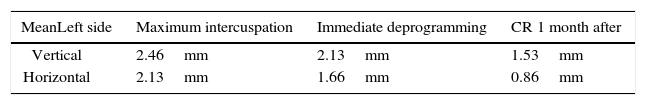
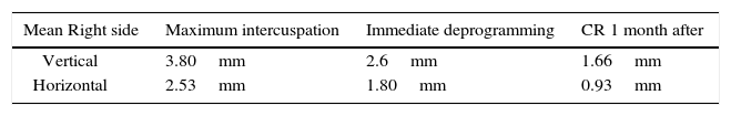
ResultsThe group of maximum intercuspation exhibited the highest mean on the right and left groups with values of ± 3.60 and ± 2.46mm in the vertical axis, while the mean for the data in the horizontal axis was ± 2.53 and ± 2.13mm, respectively. The next group was the one with the record taken with immediate deprogramming, which mean values were ± 2.60 and ± 2.13mm in the vertical axis while in the horizontal axis, the final mean was ± 1.80 y ± 1.66mm. Finally, the lowest value of displacement belonged to the group of centric relation measured 1 month after splint use. The mean values for this group were ± 1.66 and ± 1.53mm in the vertical axis and in the horizontal axis it was ± 0.93 and ± 0.86mm.
In the condylar displacement record with immediate deprogramming to centric relation a higher mean was observed on the right side in the vertical axis (± 0.94mm) and the left side in the horizontal axis (± 0.93mm). In the record of maximum intercuspation to centric relation, higher values of displacement were obtained on the right side in the vertical axis (± 2.14mm) and in the horizontal axis (± 1.60mm) compared to those of the left side (Tables IandII).
In regard to the displacement direction, 11 of 15 patients showed a posterior and downwards displacement on the right side, while on the left side 9 of 15 recorded this same pattern, describing the path normally caused by premature contact due to a dental fulcrum in the posterior area of the arch.
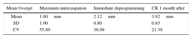
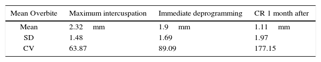
Dental shiftAs far as the results for dental shifts, the data were obtained after taking records at three different times in order to perform basic statistic tests and afterwards, carry out a clinical analysis and obtain the measurements of the horizontal and vertical axis. These values are found in tables IIIandIV, respectively.
If one chooses to follow the guidelines established in 2001 by the AAO, orthodontic treatment must involve within its goals: to achieve a correct occlusal function and to provide skeletal, dental and muscle stability and health.8 From these parameters, reducing discrepancies between maximum intercuspation and centric relation should be considered as one of the objectives in treatment planning. Such reduction, according to previous studies, yields data that could make the clinician differ in the diagnosis and treatment planning.9
The results obtained in this research were achieved after the analysis of a mounting on a semi-adjustable articulator. The centric relation record obtained with an initial immediate deprogramming that uses an anterior stop has been validated previously as an appropriate technique for recording the mandible in this position. It is assumed that when the patient wears this device, a more relaxed muscle position is achieved. However it is not correct to consider such relaxation or the soachieved position as real or stable in a patient who has previous engrams and symptoms. Many studies have shown that neuromusculature guides the position of the jaw to maximum intercuspation, regardless of the final position of the condyles. As a result, the mandibular position will be based on occlusion and erroneously considered by many clinicians as the patient’s stable condylar position. Due to the effect of the neuromusculature, mandibular manipulation becomes unreliable. Calagna et al.6 determined that there is no scientific method available to determine which patients may be exempt from neuromuscular reconditioning and that deprogramming is the key to the reproducibility of a given position.
An appropriate method for providing the highly desirable muscular-skeletal stability and ensuring condylar settlement as well as positional dental and arch changes between MI and CR, is the use of an occlusal splint for a period of time determined by clinical outcomes. Due to muscle influence it is not possible to detect the existence of occlusal premature contacts through simple visual examination or by means of trimmed models. Spear,10and subsequently Okeson,11 reported that a posterior premature contact may cause the condyle to displace the disk while the mandible works as a pivot to dodge the contact and reach maximum intercuspation. By obtaining this new posture, a more physiological diagnostic start point may be considered for treatment decisions.1
On the other hand, the effectiveness and reliability of the CPI has been previously documented as a viable method for measuring condylar slides.7
The results of the present study did not show statistically significant differences between the centric relation records obtained with immediate deprogramming and the ones subsequent to the use of an occlusal splint in regard to condylar displacement except for the left side in the horizontal axis. In terms of dental shifts, there was a statistically significant difference in the overjet but not in the overbite. However, there were significant differences in the results of the records at maximum intercuspation and at centric relation subsequent to the use of the occlusal splint, with the exception of condylar displacement of the right side on the vertical axis.
Previous studies have shown that when there is a discrepancy between maximum intercuspation and centric relation, the overjet increases while the overbite decreases. This has been demonstrated in the present study: the overjet increased on an average of ± 3.92mm while the overbite decreased by a total average of 1.11mm. Thus, these results are consistent with those described by Karl and Foley5 as well as with the ones described by Cordray.1
The results of this research obtained from symptomatic patients after articulator mounting with centric relation records subsequent to the use of an occlusal splint and compared with a position of maximum intercuspation showed premature contact points and an increase in the overjet that suggested a molar and canine Class II tendency according to Angle’s classification and a decrease in the overbite that showed an open bite tendency.
The magnitude of the centric relation discrepancy at the condyles has influence over occlusal relationships by changing the malocclusion’s type or severity, according to the mandibular position adopted during the analysis. This discrepancy cannot be measured directly in the mouth due to the different features present in each patient: facial type, goniac angle and occlusal plane angle. This means that patients with different facial characteristics displayed major or minor differences in arch relationship even in the presence of the same amount of condylar displacement. For this reason, authors such as Roth,12 Wood,7Cordray,1 Fantini,13 Girardot,14 Utt,15 Karl5 and Hidaka16 recommend orthodontic diagnosis based on models articulator-mounted in centric relation so as to be able to identify discrepancies that could be disguised.2
These differences found in inter-arch relationships and condylar position may affect different decisions in orthodontic treatment. (1) Diagnosis: magnitude (mm) of horizontal, vertical and transverse discrepancy; mandibular growth direction and the expected mandibular rotation during treatment; (2) treatment plan: extraction or non-extraction, surgical; (3) anchorage requirements; (4) treatment mechanics (indicated by the previous points); (5) occlusal finishing (arch coordination in the 3 planes of space); (6) Assessment of the effects of orthodontic treatment; and (7) Assessment of relapse.1
The majority of patients observed in this study showed posterior lower condylar displacement, which determines the presence of dental fulcrums. With a pure dental fulcrum, the primary dental contact in centric relation is usually found in the posterior teeth. While the patient tries to reach maximum intercuspation, this primary contact point works as a mandibular rotation zone. The anterior portion of the mandible will rotate counter-clockwise in order to close. The segment posterior to this contact point, which contains the condyle, rotates in a clockwise direction.
In the presence of anterior displacements, while the mandible closes toward maximum intercuspation, it also rotates forward by means of dental inclinations. The condyle cannot, therefore, move in a horizontal direction as it is positioned in the posterior eminence of the articular eminence in centric relation.
Primary contact dental shifts to maximum intercuspation show different patterns when the contact is made at a marginal ridge, a cusp or in anterior teeth or posterior teeth. In turn, the contact may be bilateral or unilateral and each contact will individually affect the condyles in the three planes of the space. Obviously it should be considered that the CPI provides unidirectional graphics data, while the actual motion of the patient is a product of a three-plane vector.
Due to the fact that dental anatomy and mandibular condyle dimensions are different for each individual, the degree of the expression of the dental shift at condylar level will be different for each patient. Therefore, since condylar movement depends on both the resulting vector as well as the individual geometry of the masticatory system, it is impossible to predict the length of a condylar shift from maximum intercuspation to centric relation by means of simple observation of the dental shift.
Of this, it can be inferred that the clinician will not be able to deduce condylar position and its changes through dental shifts. The clinician should be aided by proper, efficient and reliable auxiliary instruments.
Since it has been demonstrated that patients with small condylar displacements have larger dental shifts and vice versa, it is recommended to use an articulator to mount every case in order to effectively analyze this situation. The abovementioned phenomenon might explain why some patients with severe malocclusions have minimal or no symptoms, whereas patients with relatively normal occlusions and small dental shifts exhibit severe symptoms. Of course this reasoning does not take into consideration other factors such as stress or each patient’s capacity to adapt.3
Conclusions- a)
There were no significant differences in the results obtained from the centric relation records with immediate deprogramming and the ones in centric relation subsequent to the use of an occlusal splint.
- b)
There is a considerable discrepancy in condylar and dental displacement between a position of maximum intercuspation and centric relation subsequent to the use of an occlusal splint, as well as in the reduction of symptoms in patients with the use of this device.
- c)
Therefore, articulator mounting is recommended for diagnostic purposes as well as measurement of condylar position and use of occlusal splints in patients with pain in order to more reliably identify the goals to consider during and through orthodontic treatment. Otherwise, these might be hidden and determine a different course in treatment planning.