


INTRODUCTION
Atopic dermatitis or atopic eczema is a chronic and recurrent inflammatory disease with exacerbations triggered by different stimuli (1). Clinically, it is characterized by pruritic lesions with scaling leading to lichenification, the latter considered to be the cutaneous component of the atopic complex (2, 3).
Several atopic dermatitis patients present recurrent or persistent infections and should be submitted to immunologic evaluation (4). Studies have demonstrated some deficiencies in the immunologic response of atopic dermatitis patients; the type, degree and prevalence of these defects however are as yet unclear (5).
The diagnosis of deficient phagocytic activity by mononuclear phagocytes is less often made as compared with the diagnosis of deficient polymorphonuclear neutrophil phagocytosis, as seen in chronic granulomatous disease, in malnutrition and other nutritional disturbances (6-8). In the presence of altered mononuclear phagocytic activity, serious infections are encountered especially by fungi, but also by viruses and intracellular bacteria. Infections are caused by Candida sp, M. gypseum, T. tonsurans, Mycobacterium tuberculosis and any type of virus.
The prognosis of atopic dermatitis with immunologic involvement is directly related to early diagnosis and specific treatment, as seen in practically all immune deficiencies.
Few cases have been described in the literature of patients with atopic dermatitis who had deficient mononuclear phagocytic activity, even though this is so important for adequate treatment and evolution of the patients' disease.
CASE REPORTS
First case
G.P.M., female, 17 years old. At the age of eleven, she started to present intense pruritis over the whole body when the diagnosis of atopic dermatitis was made. At 13, there was aggravation of the lesions with diagnosis of Tinea corporis. This was so serious that she often could not go to school. At the age of 17, she presented papulo-vesicular lesions with erythematous base and serous exudate associated with whitish round lesions with definite contour, disseminated all over her body, in which state she was first seen at our Immunology Section. There was also an intense vaginal discharge.
Second case
T.A.S., male, 2 years old. Diagnosis of atopic dermatitis was made at the age of one. The clinical presentation aggravated when the atopic dermatitis developed fungal infection which responded poorly to topical treatment as well as frequent recurrences. At the age of 2, he was brought to the Immunology Section with intense pruritis, papulo-vesiculo-erythematous confluent lesions predominantly on the face, extensor regions and lower members associated with crusty lesions.
Third case
F.C.N., male, 12 years old. Diagnosis of atopic dermatitis was made at the age of three. At the age of four, he developed Cryptococcus neoformans meningitis confirmed by positive cerebrospinal fluid culture, associated with whitish oral mucosa lesions. At the age of ten, he developed crusty erythematous lesions on the face, trunk and members.
Fourth case
H.G.M., female, 12 years old. At the age of three, she presented with disseminated maculo-papular lesions, mainly on the extensor surface of the members, which progressed to lichenification. There was aggravation and at the age of four, there was alopecia with tonsured hair, areas of scaling on the face, trunk, upper members, nail thickening, labial fissures and repeated viral infections.
Fifth case
J.A.S., female, 17 years old. At the age of two, she had erythematous exudative lesions on the face. At the age of three, these lesions spread to the whole body, except hand palms and foot soles, with intense pruritis. At the age of seven, there was aggravation of her condition, when she arrived at the Immunology Section with severe atopic dermatitis with fungal infection.
RESULTS
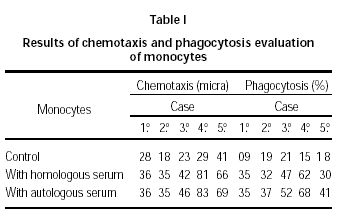
Our patients were positive for air allergens (Dermatophagoides pteronyssinus and/or farinae). Cultures of skin scales showed Candida sp, M. gypseum and dermatophyte T. tonsurans. In the first case, Candida sp was found in the vaginal discharge. In all five patients, the values of immunoglobulins, lymphocytes, T and B cells, CD4 + and CD8 + cells, C3 and C4 components of complement as well as chemotactic and phagocytic activity for neutrophils were normal while phagocytosis was persistently decreased in monocytes.
All five patients showed decreased phagocytic ingestion phase for monocytes (table I) when compared to normal values: 27 ± 9; 78 ± 7; 75 ± 10 to 27 ± 5; 65 ± 7; 68 ± 7, according to age (9).

After the diagnosis of atopic dermatitis with dermatophytosis, associated with deficient monocyte phagocytic activity was made, all patients received systemic antifungal treatment during the fungal infection. No concurrent macrolides were given and there was control of hepatic transaminases and coagulation tests. Prophylactic use of cetoconazol shampoo was also initiated. Immediately at first signs of fungal infection, systemic antifungal therapy was started once more, always with hepatic function control. The patients started to have satisfactory response to the treatment.
DISCUSSION
Zimosan (Zy) particle phagocytosis used in our methodology assesses the monocyte ingestion phase (9). Monocytes present receptors for C3b and C5b implicated in the ingestion phase of phagocytosis, where C5b is considered the more important one. Mononuclear cell incubation with serum promotes complement activation, resulting in the formation of C3b and C5b components which unite to Zimosan particles. The union of C3b and C5b with their respective receptors on the surface of monocytes culminates with the ingestion of the Zimosan particles by these phagocytes. The control test evaluates the viability of the method with spontaneous particle ingestion. The second and third tests evaluate zimosan ingestion in the presence of C3b and C5b components of heterologous and homologous serum respectively.
The results demonstrated no significant difference between the second (mononuclear leucocytes incubated with Zy and human serum "pool") and the third test (mononuclear leucocytes incubated with Zy and the patient's serum), with normal complement values of these patients (10), indicating that the decrease in phagocytosis was due to an intrinsic monocytic defect. These data are coherent with the greater frequency of fungal infections in these patients. The children studied were of normal height and weight, which makes it impossible that the phagocytic deficiency in monocytes might be due to malnutrition (9).
Studies confirm that the skin of atopic dermatitis patients is more frequently colonized by Staphylococcus aureus than non-atopic individuals and that this colonization is more intense in the presence of more serious degrees of dermatitis, which contributes to the chronicity of the disease (11, 12).
Patients with atopic dermatitis may show exacerbation of their eczema which is triggered by various inflammatory stimuli, through IgE mediated mechanism, including to dermatophytes; these patients thus become sensitized to these agents and are more susceptible to cutaneous dermatophyte infections (1, 13). The positive tricofitin reactivity observed in atopic dermatitis does not necessarily signify sensibilization to the dermatophytes but is probably a sign of cross-reaction to fungus (14).
Candida albicans which belongs to the normal human microflora, can induce synthesis of specific IgE in patients with atopic dermatitis and asthma. The Candida albicans sensibilization manifested in children with serious atopic dermatitis is frequently associated with immunodeficiency (15) and has been suggested as a component of the atopic dermatitis pathogenesis (16). On the other hand, eczema is one of the cutaneous manifestations of primary immunodeficiency which leaves a doubt as to the initial disease factor (17). Neutropenic patients have greater risk to develop systemic infections by Candida albicans, while the fungus also has the ability to activate Th2 response as evasive strategy (18, 19).
An increasing number of children with atopic dermatitis are recognized to possess immune defects, while the exact incidence is still unknown (5). Monocytes and T helper lymphocytes exert an important role in the immunologic dysfunction of atopic dermatitis. Studies have evaluated the citocin pattern in the immune response of atopic dermatitis, but results have been conflicting, while the main studies realized have used stimulation by different mitogens, which makes comparison difficult (20). Fisher et al have observed significant decrease in chemotaxis by monocytes in patients with serious mucocutaneous candidiasis and also in some patients with atopic dermatitis (21).
These observations as well as the literature propose the hypothesis of deficient mononuclear leukocyte phagocytic activity in patients with atopic dermatitis associated with fungal infections and alert that special therapeutic measures must be taken in order to improve these patients' quality of life.
