


Spontaneous rupture of hepatic metastasis causing hemoperitoneum is a rare entity. Ruptured hepatic metastasis has typical imaging findings on biphasic CT which may help in clinching the diagnosis. We present a case of rupture hepatic metastasis from choriocarcinoma in a young-female patient who was managed by transcatheter hepatic artery embolization. A brief review of the imaging features and therapeutic options for the ruptured hepatic metastases is discussed along with the case.
Choriocarcinoma is a gestational trophoblastic tumor of syncytiotrophoblastic origin. It is most commonly arises from body of uterus and has a propensity to metastasize hematogenously to lungs, vagina, ovaries and central nervous system.1 Liver metastasis is uncommon and occurs in 4-20% patients.1 Ruptured hepatic metastasis from choriocarcinoma as a cause of hemoperitoneum is a very rare entity.
Case ReportA 26-year-female presented with sudden onset pain abdomen for two days. On examination, she was pale, had tachycardia and hypotension. Her abdomen was tender to palpation. Laboratory investigations showed marked anemia (Hb-6 gm%), however other investigations including liver and renal function tests were within normal limits. Ultrasound abdomen showed multiple small (1.5to 2 cm) heterogeneously hyperechoic focal lesions in both the lobes of liver with a large subcapsular collection and a small amount of free fluid in the abdomen. Small echogenic (atleast three) lesions were seen in the spleen also. In addition, a large heterogeneous lesion was also seen in the pelvis from which uterus and ovaries were not seen separately. Biphasic CECT abdomen showed multiple small hypervascular lesions in both lobes of liver some of them located peripherally in subcapsular location with discontinuity in hepatic surface (Figure 1). Similar lesions were also seen in the spleen. There was a large hypervascular lesion with areas of necrosis in the pelvis from which the uterus and ovaries were not seen separately (Figure 2). Multiple dilated and tortuous vascular channels were seen with in the pelvic wall. There was mild free fluid in the abdomen. With provisional diagnosis of ruptured hypervascular metastases likely from choriocarcinoma, transcatheter hepatic artery embolization was done with gel foam and polyvinyl alcohol (500-700 μ) particles (Figure 3). Selective splenic artery angiogram also showed similar three lesions. Serum beta-hCG levels were done which was elevated (8,400 IU/mL). The general condition of the patient improved temporarily and ultrasound abdomen showed reduction in size of the subcapsular hematoma. However on the sixth day of admission patient developed left hemiparesis and head CT showed a large haemorrhagic lesion in the right frontal lobe. USG guided FNAC from the liver lesion showed metastatic choriocarcinoma (Figure 4). Patient developed bleeding per vaginum on the same day for which vaginal packing was done. Chemotherapy for choriocarcinoma could not be started due to poor general condition of the patient. On the ninth day, due to continued bleeding per vaginum uterine artery embolization was done with gel foam and polyvinyl alcohol (500-700 μ) particles. However the patient continued to deteriorate and succumbed to the disease on the eleventh day of admission.
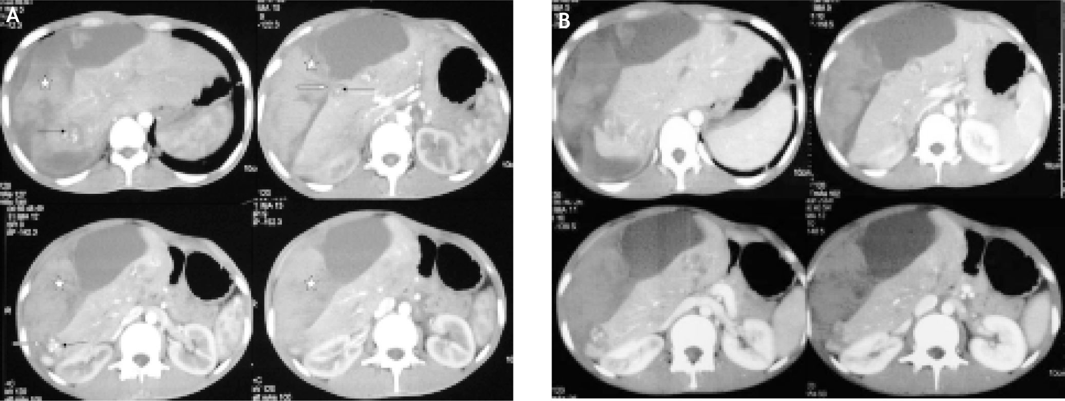
 and venous (B) phase showing multiple small hypervascular lesions (black arrows) with relative washout in portal venous phase in both lobes of liver, mostly located peripherally in subcapsular location and few of the lesions showing discontinuity at hepatic surface (white arrows). Large heterogeneously hyperdense subcapsular collection with hematoma in right subphrenic region (asterix) is also seen.")
Biphasic CECT abdomen in arterial (A) and venous (B) phase showing multiple small hypervascular lesions (black arrows) with relative washout in portal venous phase in both lobes of liver, mostly located peripherally in subcapsular location and few of the lesions showing discontinuity at hepatic surface (white arrows). Large heterogeneously hyperdense subcapsular collection with hematoma in right subphrenic region (asterix) is also seen.

 and contrast blush with persistence of the blush in the venous phase. Post-embolization angiogram showing complete occlusion of the hepatic artery.")
Selective hepatic artery proper angiogram revealed multiple abnormal tortuous vessels in both lobes of liver with aneurysmal dilatation of their ends (black arrows) and contrast blush with persistence of the blush in the venous phase. Post-embolization angiogram showing complete occlusion of the hepatic artery.

Non-traumatic spontaneous hepatic hemorrhage is most commonly caused by ruptured hepatocellular carcinoma or hepatic adenoma.2,3 The other liver tumors which are likely to cause hemorrhage are focal nodular hyperplasia, hemangioma and metastasis.2 Spontaneous rupture of hepatic metastasis has been described in metastasis from lung, pancreas, stomach, kidney, testis, breast, prostate, gall bladder, melanoma and choriocarcinoma.4-6 Rupture of a metastatic liver tumor is rare as they tend to be more fibrotic, less vascular and invasive, and have lesser propensities to penetrate the liver capsule as compared to hepatocellular carcinoma.7 However, choriocarcinoma being a vascular and hemorrhagic tumor has a propensity to rupture especially when in subcapsular location due to direct pressure by the tumor against the capsular surface because of increased intra abdominal pressure.5 Other mechanisms of rupture that have been described are necrosis and breakdown of a tumor nodule or increased intravascular pressure secondary to tumor emboli, resulting in intra hepatic venous obstruction.4
The imaging appearance of choriocarcinoma metastasis is like other hypervascular lesions. On ultrasound, there are usually multiple hyperechoic lesions. Biphasic CT imaging shows the lesions to be hypervascular in arterial phase with washout in portal venous phase. Rupture is suspected when there are hemorrhagic lesions with subcapsular hematoma in relation to the hepatic lesions with or without hemoperitoneum. MRI also shows similar hyper vascular lesions. Angiographic findings include a hypervascular mass with aneurysmal dilatation of the peripheral end of the hepatic arteries (grape like appearance) at the arterial phase and persistent vascular lakes at the venous phase.5,6 The helpful diagnostic indicators of ruptured hepatocellular carcinoma are peripheral location, protruding contour, discontinuity of the hepatic surface, and surrounding hemoperitoneum.3
Treatment of spontaneous rupture of choriocarcinoma metastasis of liver depends on the tumor size, tumor location, and severity of bleeding.7 Surgery is usually difficult in these patients due to hemodynamic instability, considerable difficulty in achieving local hemostasis by suture ligature or packing with topical haemostatic agents and hepatic resection is usually not possible due to multiple lesions.8 The best alternative is transcatheter hepatic artery embolization (TAE) which is a palliative therapeutic option for other causes of bleeding hepatic metastases, including those with massive hemorrhage caused by spontaneous rupture. TAE should be selected to control the bleeding in these cases because the primary tumour is extrahepatic in location and surgical resection is not curative. Prognosis for patients with ruptured choriocarcinoma hepatic metastasis is poor. They usually have extensive metastasis elsewhere and spontaneous rupture of liver metastasis is usually a terminal event. Mean survival is usually less than 6 months and in most less than 6 weeks.7
To conclude, rupture of choriocarcinoma metastasis is very rare entity. This diagnosis should also be considered besides hepatic adenoma, when a young female patient in reproductive age group presents with acute abdominal pain and liver lesions. In our case, the hepatic lesions showed atypical hypervascularity along with the complex pelvic lesions that hinted the diagnosis of choriocarcinoma which was confirmed by hCG levels and FNAC.


