


Patients with metabolic dysfunction-associated steatotic liver disease (MASLD) are at an increased cardiovascular risk. On the contrary, non-alcoholic fatty liver disease (NAFLD) is highly prevalent in patients with coronary heart disease (CHD). However, it is not known whether patients with significant CHD show a higher frequency of liver fibrosis. This study aimed to determine the frequency of MASLD and liver fibrosis in patients with CHD and to assess whether coronary stenosis is significantly associated with MASLD and fibrosis.
Patients and MethodsThis observational and analytical study included adult patients without any known liver disease who underwent coronary angiography for suspected coronary artery disease (Jul 2021–Jul 2022). The presence of significant CHD (> 50 % stenosis of at least one coronary artery) was determined. Liver elastography (FibroScan®) was performed up to 6 months after the coronary angiographic study to determine liver fibrosis, a measurement of liver stiffness (> 6.5 Kpa). Fisher's test, Mann–Whitney U test, and logistic regression models were used (p < 0.05).
ResultsThe study included 113 patients (76 % men, average age: 63 years [standard deviation: 9.9]), of which 72 % presented with significant CHD. The prevalence rate of MASLD was 52 %. Liver fibrosis was present in 12 % of the patients and all patients in the significant CHD group (p = 0.007). An increase in the number of vessels with significant CHD increased the probability of liver fibrosis (odds ratio, 1.79; 95 % confidence interval, 1.06–3.04; p = 0.029).
ConclusionsMASLD is highly prevalent in patients with significant CHD but without known liver damage. These data suggest that MASLD and liver fibrosis should be investigated in patients with CHD. The presence of confounding variables, especially the presence of type 2 diabetes mellitus, should be evaluated in further studies.
Metabolic dysfunction-associated steatotic liver disease (MASLD) is the most common cause of chronic liver disease worldwide, with an estimated prevalence of 25 %. There are regional and ethnic differences, such as an estimated 31 % prevalence in South America [1]. In Chile, data from 2009 showed a prevalence of 23 % in the general population, which is estimated to be increasing [2]. The high prevalence of MASLD is strongly associated with metabolic syndrome and the increase in the rate of obesity. Chile is currently the country with the highest obesity rate, reaching 34.4 % of the population aged over 15 years [3].
Patients with MASLD have been shown to have an increased 10-year risk of cardiovascular events compared with healthy individuals. Cardiovascular disease is the leading cause of death in patients with non-alcoholic fatty liver disease (NAFLD) [4]. Moreover, patients in advanced stages of NAFLD and with liver fibrosis present with a higher rate of fatal and non-fatal cardiovascular complications, which are associated with the severity of fibrosis [5–7]. Thus, the current literature recognizes the relationship between NAFLD and coronary heart disease (CHD) [8,9].
Conversely, an increased risk of NAFLD has been reported in patients with CHD. Previous studies have shown a higher prevalence of NAFLD in patients with CHD, the frequency of which increased with the severity of CHD [10]. However, this “dose-response” between NAFLD and CHD severity was questioned in subsequent studies [8]. Moreover, evidence supporting NAFLD as a risk factor for CHD is scarce and controversial.
This possible bidirectional relationship between NAFLD and CHD could be due to the common risk factors for both diseases. In a recent sub-analysis of the Framingham study, the presence of liver fibrosis was found to be associated with multiple cardiovascular risk factors, which could explain the higher cardiovascular mortality in patients with NAFLD [9]. Consequently, the current question in the literature is whether these cardiovascular risk factors are responsible for the association between NAFLD and CHD or whether liver fibrosis is an independent cardiovascular risk factor. The prevalence of liver fibrosis in patients with CHD is currently not known.
This study aimed to determine the frequency of MASLD and liver fibrosis in patients with CHD and to assess whether significant coronary stenosis is associated with steatosis and liver fibrosis.
2Material and methods2.1Design and study populationThis observational, cross-sectional study was conducted in adult patients who underwent coronary angiography for suspected coronary artery disease between July 2021 and July 2022 at the Hospital Clínico Universidad de Chile. Patients with an indication for coronary angiography for non-ischemic causes, pregnant women, patients with transaminases greater than or equal to five times the upper limit of normal, history of known chronic liver damage, personal history of cancer, abdominal surgery up to 3 months prior, and history of heart failure or cardiac hypodebit syndrome (defined as left ventricular ejection fraction <50 %, as measured by transthoracic echocardiography or ventriculography) were excluded.
2.2Clinical information, exposure and outcomesPatients who had undergone coronary angiography were telephonically invited to participate in the study and selected consecutively according to the inclusion and exclusion criteria during the study period.
Trained personnel collected demographic data, clinical history, and laboratory test reports (closest within six months of the date of coronary angiography) from the clinical records using an established form. Coronary angiography revealed the number of vessels affected and the magnitude of the stenosis. Significant coronary heart disease is defined as the presence of > 50 % stenosis in at least one coronary artery [11]. Hepatic steatosis and fibrosis were determined through liver elastography (Fibroscan® model 530 Compact 2018); it was performed up to 6 months after coronary angiography by an expert operator. According to previous evidence, hepatic steatosis was defined as the presence of > 5 % hepatic fat (controlled attenuation parameter [CAP] > 237 dB/m) and significant fibrosis was determined at the liver stiffness measurement (LSM) of ≥ 6.5 kPa [12]. Alcohol consumption was considered as > 3 drinks/week in men and > 2 drinks/week in women.
2.3Statistical analysisA sample size calculation was performed according to the literature-estimated prevalence of NAFLD of 31 % [1], with a 95 % confidence interval (CI) and a precision of 10 %, resulting in 91 patients (considering a loss-adjusted sample of 10 %).
The normal distribution of continuous variables was assessed using the Shapiro–Wilk test. Descriptive statistics were expressed as frequency and percentage for categorical variables and as mean (standard deviation) or median (interquartile range [IQR]) for continuous variables, as considered appropriate. Bivariate analysis was performed using Student's t-test or Mann–Whitney U test, as considered appropriate (for continuous variables), and Fisher's exact test (for categorical variables).
To test the association between significant coronary stenosis and hepatic fibrosis, we considered the exposure in two different manners: estimating its linear effect on the outcome by using the absolute number of vessels with significant CHD, and as a dichotomized variable by grouping the patients in a group with two or more vessels with significant CHD and other with one or less vessels with CHD. Thus, different logistic regression models were performed to evaluate the effect of significant CHD (independent variable) on hepatic fibrosis (dependent variable) adjusted by the effect of T2DM and obesity in separate models. For each variable included in the models, we calculated the odds ratio (OR) with its 95 % confidence interval (CI) and p-values. All analyses were performed using Stata 14.0 (StataCorp. 2015. Stata Statistical Software: Release 14. College Station, TX, StataCorp LP) software. p-value <0.05 was considered strong evidence against the null statistical hypothesis.
2.4Ethical statementsWritten informed consent was obtained from each patient included in the study, and the study protocol conforms to the ethical guidelines of the 1975 Declaration of Helsinki as reflected in a priori approval by the Ethics Committee of Hospital Clínico Universidad de Chile, Santiago, Chile (No. 33/2021).
3Results3.1Patient characteristicsDuring the study period, 702 coronary angiographies were performed, of which 113 patients met the inclusion criteria, 81 (72 %) of whom presented with significant CHD on coronary angiography. Of the patients without significant CHD (n = 32), 24 had normal coronary angiography with no lesions; the remaining eight patients had less than 50 % stenosis in at least one artery. There were no differences between the latter two groups (normal coronary angiography vs. less than 50 % stenosis) in terms of demographic variables (age, sex), clinical variables (hypertension or diabetes mellitus 2), anthropometry, or steatosis. None of the patients had fibrosis.
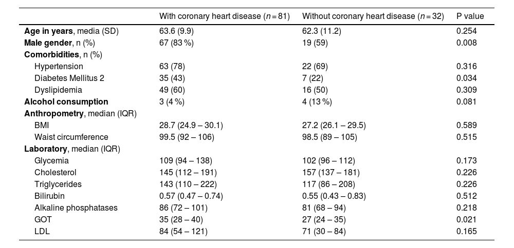
Table 1 describes the baseline characteristics of the patients, highlighting a higher proportion of male patients with CHD than those without (83% vs. 59 %, p = 0.008) and a greater presence of type 2 diabetes mellitus (T2DM) in the same group (43% vs. 22 %, p = 0.034). Furthermore, T2DM increased the odds of having two or more vessels affected by CHD (OR = 3.1 [CI 95 %: 1.4 - 6.9], p-value = 0.006. In addition, glutamic-oxaloacetic transaminase levels were higher in the CHD group than in the group without CHD (median, 35 U/L [IQR 14–176 U/L] vs. 27 U/L [IQR 17–59 U/L]; p = 0.021).
Demographic and clinical characteristics of the sample and their association with coronary heart disease.
SD, standard deviation; BMI, body mass index; IQR, interquartile range; GOT, glutamic oxaloacetic transaminase; LDL, low-density lipoprotein.
The overall frequency of hepatic steatosis was 52 %, with no significant difference between the groups with and without CHD (50.6% vs. 56.3 %; p = 0.371). The frequency of hepatic fibrosis was 12 % and was observed only in the CHD group (17.3% vs. 0 %; p = 0.010). There was no significant difference in median liver stiffness between the groups with and without CHD (4.5 [2.8–37.9] vs 4.5 [2.5–6.1] kPa, p = 0.292) (Table 2).
Steatosis and fibrosis measured by hepatic elastography and its relationship with coronary heart disease.
CAP, controlled attenuation parameter.
Patients with T2DM showed a higher frequency of liver fibrosis than those without (31% vs. 7 %, p = 0.006).
3.3Hepatic fibrosis and the number of vessels with significant coronary heart diseaseA higher frequency of hepatic fibrosis was found in patients with a higher number of vessels with significant CHD (Fig. 1). Thus, the number of vessels involved increased the probability of hepatic fibrosis (OR, 1.79; 95 % CI, 1.06–3.04; p = 0.029). When categorizing the variable into two or more affected vessels, an increased strength of association with liver fibrosis was observed (OR, 3.44; 95 % CI, 1.07–11.08, p = 0.038). However, the statistical evidence for both associations decreased when controlled for the history of diabetes mellitus (Table 3). In contrast, the number of coronary vessels affected, as linear effect or dichotomized in two or more affected vessels, remained as risk factors for hepatic fibrosis after control by obesity (Table 3). There was no significant difference in the frequency of liver fibrosis between patients with and without a history of acute myocardial infarction (11% vs. 14 %, p = 0.775).

Logistic regression analysis to estimate the risk of hepatic fibrosis according to the number of coronary vessels affected.
In this cohort of adult patients who underwent coronary angiography for suspected coronary artery disease, a high frequency of global hepatic steatosis, measured by CAP (52 %), was observed. In addition, an association between the number of vessels with CHD and the presence of hepatic fibrosis was also observed, with T2DM being a confounding factor in this relationship because it was associated with the number of vessels with CHD (the exposure) and hepatic fibrosis (the outcome).
Our results on hepatic steatosis showed a prevalence higher than that reported in the literature. In local studies conducted more than a decade ago, a 23 % prevalence of hepatic steatosis was reported [2]. However, recent international literature shows a 31 % prevalence of this disease in South America [13]. This could be attributed to the known increase in obesity and metabolic syndrome in the general Chilean population in recent decades [3]. However, the prevalence of steatosis did not significantly differ between the groups with and without CHD (51% vs. 56 %), possibly because of lack of differences in body mass index (BMI) or waist circumference between the groups. In contrast, liver fibrosis was observed in 17 % of the patients who underwent coronary angiography, with 6.2 % (5/81) having advanced fibrosis (F3–F4). Interestingly, fibrosis was only observed in the group of patients with CHD and was not found in those without CHD.
Some studies have evaluated the impact of steatosis or fibrosis on cardiovascular disease with controversial results. Ciardullo published a study that included 2734 participants and reported an NAFLD prevalence of 48.6 %, with a presence of liver fibrosis in 9.7 % [8]. This study revealed that patients with cardiovascular disease (defined as a history of CHD, and/or stroke or transient ischemic attack) had a higher incidence of hepatic steatosis (59.6% vs. 47.1 %, p = 0.013) but not fibrosis (12.9% vs. 9.3 %, p = 0.123). Similar results were observed by Choi et al., who found a higher prevalence of NAFLD in patients with coronary stenosis, as evidenced by coronary angiography (78% vs. 51 %) [10]. Conversely, in a meta-analysis evaluating the impact of liver disease on cardiovascular disease, Wen et al. observed that the incidence of cardiovascular disease in the NAFLD group was twice that in the control group (RR, 2.26; p < 0.01) [14].
Multiple studies have attempted to evaluate whether hepatic fibrosis is an independent cardiovascular risk factor or if other factors interfere with this association. In our study, we observed that the number of compromised coronary vessels increased the probability of hepatic fibrosis; however, this association was nullified after controlling for DM (present in 43 % of the patients with CHD). The above argument favors the difficulty in evaluating the causal relationship between CHD and hepatic steatosis, with or without the presence of fibrosis, owing to the multiple confounding factors that interfere with the relationship. Previous studies have described the factors associated with the presence of liver fibrosis in patients with CHD. In a recent sub-analysis of the Framingham study, which included 3276 participants, an overall prevalence of steatosis of 28.8 % and fibrosis of 8.8 % (defined as an LSM ≥ 8.2 Kpa) was observed.9 It was further observed that compared with the participants without fibrosis, those with hepatic fibrosis had a higher frequency of obesity (OR, 3.11), hepatic steatosis (OR, 3.66), metabolic syndrome (OR, 2.80), and T2DM (OR, 2.67), which remained significant after adjusting for CAP and BMI. In contrast, Ciardullo et al. did not observe an independent association between liver fibrosis or steatosis with cardiovascular disease and heart failure after adjusting the models for confounding variables, such as diabetes, smoking, and age [8].
In our study, a higher frequency of patients with T2DM was observed in the groups with liver fibrosis, and also in those with coronary artery disease, so that T2DM represents a confounding factor. However, it is important to take into consideration this subgroup of patients, since it has been proposed that patients with early stage liver fibrosis (F1) with associated T2DM have an increased risk of more severe liver disease (cirrhosis and hepatocarcinoma) or also called "rapid progressors" [15] Therefore, diabetes patients with CHD and fibrosis may have a more rapid progression of liver disease.
A recent meta-analysis investigated the incidence of cardiovascular disease and mortality in patients with MAFLD. Ten cohort studies were included in this meta-analysis, and the incidence of cardiovascular disease in the MAFLD group was found to be more than twice that in the control group (RR, 2.26; p < 0.01). Furthermore, cardiovascular disease mortality was 1.57 times higher in the MAFLD group than in the control group (RR, 1.57; p < 0.01) [14]. Although our study did not find a higher frequency of hepatic steatosis and MASLD in patients with altered coronary angiography findings, their association with hepatic fibrosis may be related to the results of this meta-analysis.
3.5LimitationsThe observational and cross-sectional design of this study limits the establishment of a causal relationship between CHD and liver fibrosis. In addition, the number of patients with liver fibrosis in this study was small, which prevented us from controlling for additional confounding factors that would have been possible with a larger sample size. However, the evaluation of steatosis and hepatic fibrosis was performed in the months following coronary angiography, a time during which patients could have made lifestyle changes, considering the recent diagnosis of CHD, thereby impacting their levels of hepatic steatosis. Also, the evaluation of hepatic steatosis and fibrosis was not performed through liver biopsy, considered the gold standard; transitional liver elastography (Fibroscan®), however, has a high diagnostic yield compared with liver biopsy. Finally, it is important to mention that we performed an objective assessment of coronary artery disease using coronary angiography rather than the noninvasive methods used in other studies, such as cardiovascular risk factors, which in turn allowed us to have a well-established control group. Thus, our research is one among the few investigations worldwide that have studied the presence of hepatic steatosis and the degree of associated fibrosis, specifically in the population with proven coronary artery disease, using validated clinical diagnostic methods.
4ConclusionsMASLD is highly prevalent in patients with significant CHD without a history of known liver damage. Although the presence of confounding variables, especially T2DM, should be evaluated in future studies, these data suggest the need for a targeted search for MAFLD and liver fibrosis in patients with CHD, especially in those with T2DM.
FundingThis research did not receive any specific grant from funding agencies in the public, commercial, or not-for-profit sectors.
Author contributionsParticipant enrollment: Luis Vega, Daniela Simian, Marcelo Salinas, Andrea Urra, Rosario Pino, Katherine Rojas. Data collection: Luis Vega, Daniela Simian, Marcelo Salinas, Andrea Urra. Statistical analysis: Abraham I. Gajardo, Daniela Simian. Results discussion: Luis Vega, Daniela Simian, Abraham I. Gajardo, Máximo Cattaneo, Juan P. Roblero, Álvaro Urzúa, Jaime Poniachik. Manuscript preparation: Luis Vega, Daniela Simian, Jaime Poniachik. Manuscript review and final approval: Luis Vega, Daniela Simian, Abraham I. Gajardo, Marcelo Salinas, Andrea Urra, Máximo Cattaneo, Rosario Pino, Juan P. Roblero, Álvaro Urzúa, Katherine Rojas, Jaime Poniachik. Jaime Poniachik is responsible for the integrity of the work as a whole.


