metricas
covid
Buscar en
Archivos de la Sociedad Española de Oftalmología (English Edition)
Toda la web
Inicio Archivos de la Sociedad Española de Oftalmología (English Edition) A new OCT sign of invasive squamous cell carcinoma of the cornea
Journal Information
Vol. 91. Issue 2.
Pages 90-93 (February 2016)
Share
Share
Download PDF
More article options
Visits
665
Vol. 91. Issue 2.
Pages 90-93 (February 2016)
Short communication
A new OCT sign of invasive squamous cell carcinoma of the cornea
Carcinoma de células escamosas en la córnea: un nuevo signo de invasión corneal en la tomografía de coherencia óptica
Visits
665
P. Rodríguez-Ausína,
Corresponding author
pazrausin@telefonica.net

Corresponding author.
, C. Hita-Antóna, C. Míguez-Garciaa, D. Antolín-Garciaa, J. Suárez-Aguadob
a Departamento de Oftalmología, Hospital Universitario de Torrejón, Torrejón de Ardoz, Madrid, Spain
b Anatomía Patológica, Hospital Universitario de Torrejón, Torrejón de Ardoz, Madrid, Spain
This item has received
Article information
Abstract
Full Text
Bibliography
Download PDF
Statistics
Figures (2)
Abstract
Case report

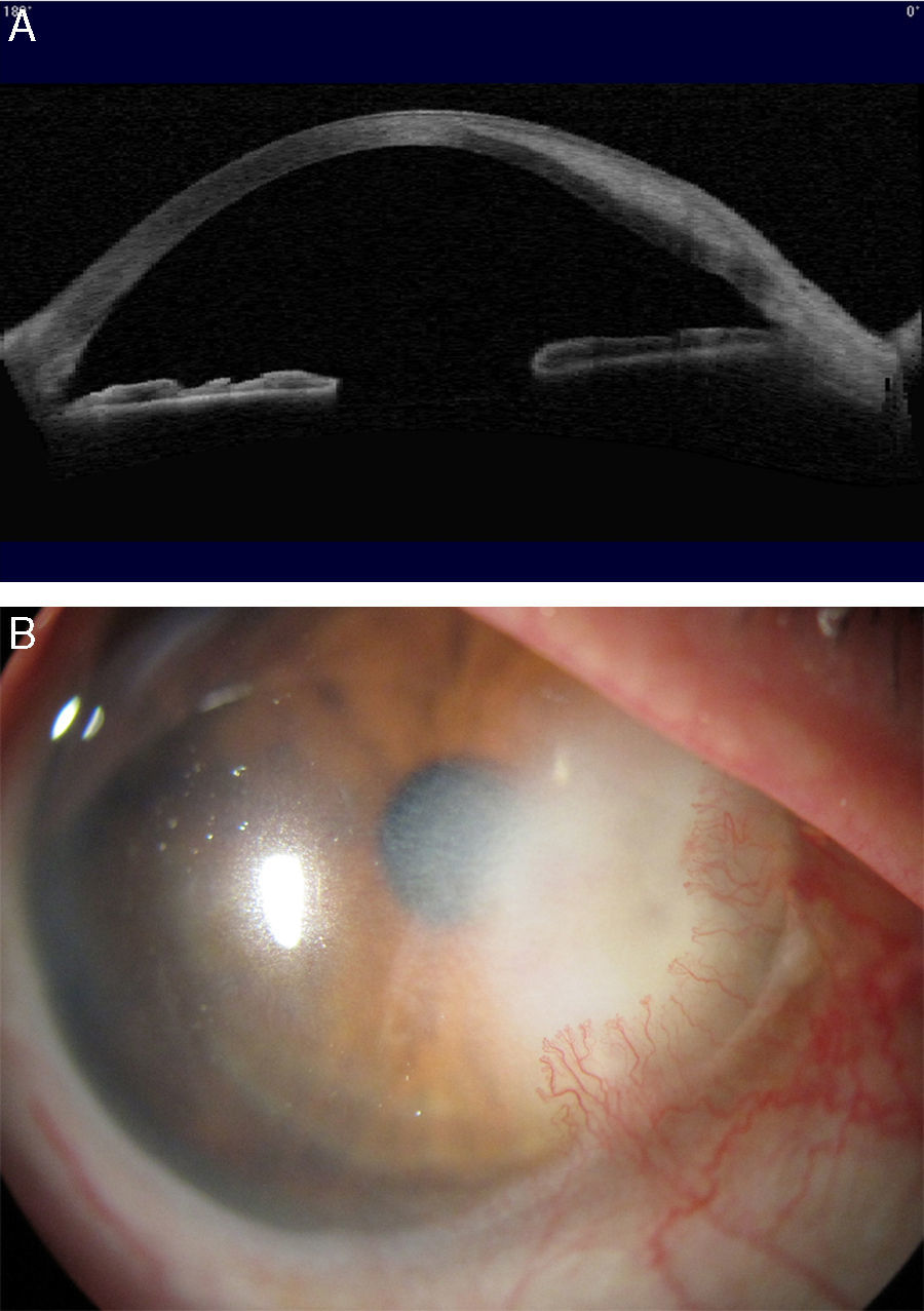
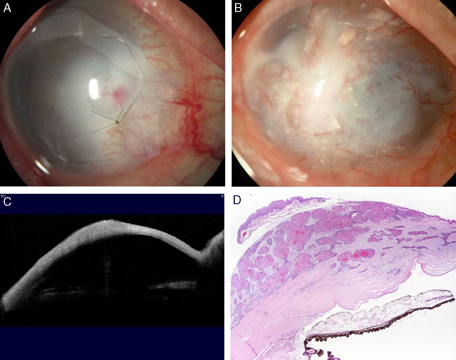
A case is reported of an unusual progressive corneal opacification and neovascularization caused by a squamous cell carcinoma (SCC) of the cornea. A patient with a white stromal infiltrate, consistent with herpetic stromal keratitis, showed a very particular image in optical coherence tomography (OCT), resembling a “tongue of lava” sliding between corneal lamellae. Histopathological analysis confirmed the diagnosis of SCC.

Discussion

To our knowledge this is the first report in the literature of this peculiar image with OCT. Squamous cell carcinoma is an extremely rare cause of progressive corneal opacification and neovascularization, and a delayed diagnosis may lead to unsuccessful treatment and loss of the eyeball.

Keywords:
Squamous cell corneal carcinoma
Stromal keratitis
Corneal infiltrate
Corneal neovascularization
Resumen
Caso clínico

Presentamos un caso de inusual opacificación corneal y neovascularización progresiva, causada por un carcinoma de células escamosas (CCE). El infiltrado estromal blanquecino sugestivo de queratitis estromal herpética, mostró una imagen en la tomografía de coherencia óptica (OCT) semejante a una «lengua de lava» deslizándose entre las lamelas corneales. El análisis histopatológico confirmó que se trataba de un CCE.

Discusión

A nuestro conocimiento, es la primera descripción en la literatura de esta peculiar imagen. El CCE es una causa extremadamente infrecuente de opacificación corneal progresiva y neovascularización, y el retraso en el diagnóstico y tratamiento puede conducir a fracaso del tratamiento y pérdida del globo ocular.

Palabras clave:
Carcinoma corneal de células escamosas
Queratitis estromal
Infiltrado corneal
Neovascularización corneal

Article

These are the options to access the full texts of the publication Archivos de la Sociedad Española de Oftalmología (English Edition)
Subscriber
Subscriber

If you already have your login data, please click here .

If you have forgotten your password you can you can recover it by clicking here and selecting the option “I have forgotten my password”
Subscribe
Subscribe to

Archivos de la Sociedad Española de Oftalmología (English Edition)

Purchase
Purchase article

Purchasing article the PDF version will be downloaded

Price 19.34 €

Purchase now
Contact
Phone for subscriptions and reporting of errors
From Monday to Friday from 9 a.m. to 6 p.m. (GMT + 1) except for the months of July and August which will be from 9 a.m. to 3 p.m.
Calls from Spain
932 415 960
Calls from outside Spain
+34 932 415 960
E-mail
Article options
es en pt

¿Es usted profesional sanitario apto para prescribir o dispensar medicamentos?

Are you a health professional able to prescribe or dispense drugs?

Você é um profissional de saúde habilitado a prescrever ou dispensar medicamentos

Quizás le interese:
10.1016/j.oftale.2023.06.012
No mostrar más