El cáncer pulmonar (CP) es el tumor con mayor frecuencia y mortalidad a nivel mundial. Casos de metástasis coroideas y retinopatía asociada a cáncer han sido publicados en CP, sin embargo no existen estudios en población mexicana que describan las posibles alteraciones retinocoroideas y su relación con el estadio de CP.
ObjetivoEvaluar a pacientes con CP para determinar la presencia de alteraciones en el segmento posterior y su relación con el estadio del mismo.
Materiales y métodosEstudio transversal y descriptivo de 50 pacientes (100 ojos) con CP. Datos demográficos: edad, sexo, tipo histológico, tiempo de evolución, estadio, tratamiento y comorbilidades. Variables de medición: agudeza visual (LogMAR), biomicroscopía del segmento anterior, registro fotográfico de retina, fluorangiografía retiniana, tomografía de coherencia óptica y electrorretinograma. Cada paciente fue evaluado por dos oftalmólogos.
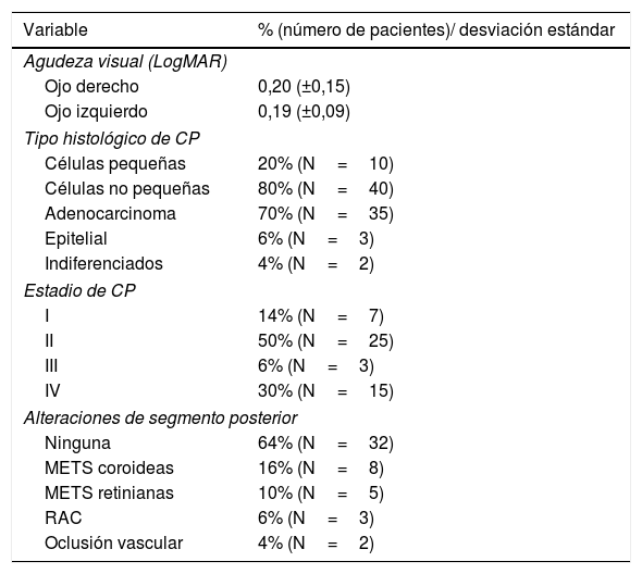
ResultadosUn total de 26 hombres y 24 mujeres fueron evaluados, el promedio de edad fue de 65 años, el tiempo medio del diagnóstico de CP fue de 6 meses siendo el adenocarcinoma el principal tipo histológico (70%), al momento de la evaluación 50% presentaban estadio II y 30% estadio IV. Las alteraciones del segmento posterior encontradas fueron: metástasis coroideas (16%), metástasis retinianas (10%), retinopatía asociada a cáncer (6%) y oclusiones vasculares (4%). La mayoría de los pacientes con alteraciones retinocoroideas se encontraban en estadio IV.
ConclusionesEn el CP pueden encontrarse oclusiones vasculares, retinopatía asociada a cáncer y metástasis a coroides y retina con una incidencia mayor a la publicada en la literatura, siendo más frecuentes en estadios avanzados de la enfermedad aunque pueden encontrarse desde el estadio II en pacientes asintomáticos.
Lung cancer (LC) is the most common tumour, and the leading cause of cancer-related death worldwide. Although cases of choroidal metastasis and cancer-associated retinopathy have been reported in LC, no studies have been conducted on the Mexican population to describe retinochoroidal findings during the course of LC, and the relationship with its stage.
ObjectiveTo evaluate patients with a diagnosis of LC, and to describe the posterior segment findings in relationship to the stage of LC.
Materials and methodsA cross-sectional and descriptive study was conducted on 50 patients with LC (100 eyes). The demographic data included age, gender, histological type, evolution time, stage, treatment, and comorbidities. The measurement variables included visual acuity (LogMAR), anterior segment biomicroscopy, retinal photography, fluorescein retinal angiography, optical coherence tomography, and electroretinogram. All patients were evaluated by two ophthalmologists.
ResultsThe study included a total of 26 men and 24 women, with a mean age of 65 years, and a mean time from LC diagnosis of 6 months. The principal histological type was adenocarcinoma (70%), and most (50%) were in stage II at the time of evaluation, with 15 (30%) patients having a metastasis (stage IV). The changes in the posterior segment included choroidal metastasis (16%), retinal metastasis (10%), cancer-associated retinopathy (6%), and vascular occlusions (4%). The majority of patients who presented with posterior segment alterations were in stage IV.
ConclusionsVascular occlusions, cancer-associated retinopathy, choroidal and retinal metastases may be found in LC, with an incidence higher than that reported in the literature, especially in advanced stages of LC, although they can be found from stage II in asymptomatic patients.
Artículo
Comprando el artículo el PDF del mismo podrá ser descargado
Precio 19,34 €
Comprar ahora


