


The epsilon toxin, produced by Clostridium perfringens, is responsible for enterotoxemia in ruminants and is a potential bioterrorism agent. In the present study, 15 regions of the toxin were recognized by antibodies present in the serum, with different immunodominance scales, and may be antigen determinants that can be used to formulate subunit vaccines.
The epsilon toxin (ETX), produced by Clostridium perfringens types B and D, is responsible for enterotoxemias in domestic ruminants, leading to great economic loss.1 In addition, ETX is considered a potential bioterrorism agent.2,3 Despite the recognized importance of this toxin, epitopes mapping studies of ETX are scarce.4 These studies can contribute to identifying interaction sites between the toxin and its neutralizing antibodies, leading to a better understanding of its neutralizing mechanisms, thus facilitating the development of new vaccines and treatments against ETX.4 The present study aimed to map continuous epitopes at the ETX by using the SPOT-synthesis technique followed by enzyme immunoassays using hyperimmune serum against the purified toxin.5
ETX was produced and purified according to the method described by Parreiras et al.6 Being the main producer of ETX and main causer of disease, C. perfringens type D strain was fermented in a bench bioreactor (BioFlo 110, New Brunswick Scientific C.O., UK) and concentrated 10 times using an ultrafiltration system with a 10kDa membrane (Amicon, Beverly, USA). The toxin was purified by ion exchange chromatography (DEAE-Sepharose CL6B).
The purified ETX was subjected to inactivation by the addition of 0.5% formalin (v/v). Aluminum hydroxide [Al(OH)3] was added as an adjuvant to obtain a final concentration of 2.83% of free aluminum oxide (Al2O3).7 Three New Zealand rabbits weighing between 1.5 and 2.5kg were given five subcutaneous inoculations with 100μg of the epsilon toxoid. The animals were immunized on days 0, 28, 49, 70 and 91, and total blood collection was performed on day 105. The serum obtained was titrated by an indirect Enzyme-Linked Immunosorbent Assay (ELISA) according to the method described by Chavez-Olortegui et al.8 A total of 100μL of a solution containing 2μg/mL of the previously obtained recombinant ETX toxin9 was used to coat the plates.
The parallel synthesis of peptides on a cellulose membrane was performed using the SPOT technique.10 Overlapping 15-amino acid peptides were synthesized, with changes in the three initial residues, covering the entire primary structure of ETX. The peptides were synthesized using the Fmoc-synthesis technique11 adapted for the cellulose membrane, and approximately 40 nanomoles of peptide per point in the membrane was obtained.10 The peptide synthesis was performed in an automatic synthesizer (ResPepSL/Automatic Spot Synthesizer, Intavis GmbH, Koln, Germany).
In total, 130 peptides were synthesized (Fig. 1). Peptides 107 through 130 were derived from regions that are part of the original structure of the toxin and included mutations in one or two amino acid residues. These peptides were used in the present study to evaluate the antigenic importance of mutated amino acid residues.
 and color intensity (no reactivity – no color, more reactive – darker) scales is shown. Mutated residues marked.")
ETX SPOT membrane immunochemical assay against anti-ETX hyperimmune serum. A list of the synthesized peptides and the mean reactivity of positive spots with numerical (0–5) and color intensity (no reactivity – no color, more reactive – darker) scales is shown. Mutated residues marked.
The membrane with synthesized peptides was blocked overnight in a solution containing 3% bovine serum albumin (BSA) (ID Bio, France) and 5% sucrose (Dinâmica, São Paulo, Brazil) in 0.1% Tris-buffered saline (TBS). Subsequently, the membrane was incubated with the hyperimmune serum diluted in blocking solution. A dilution was used for which the absorbance in the indirect ELISA was close to 1.0.5
Binding between antibodies present in the serum and the peptides was detected by incubating the membrane with rabbit anti-IgG conjugated with alkaline phosphatase at the dilution recommended by the manufacturer (Sigma–Aldrich, St. Louis, USA). The substrates used consisted of 3-(4,5-dimethylthiazol-2-yl)-2,5-diphenyltetrazolium bromide (MTT), 5-bromo-4-chloro-3-indolyl phosphate (BCIP) and MgCl2 (Sigma). After 20min, the reaction was stopped by discarding the reagents and washing the membrane with distilled water. The protocol described above was repeated twice with the hyperimmune serum and once with preimmune rabbit serum.
During the immunochemical assays, the anti-ETX hyperimmune serum was able to bind to some peptides and to detect the antigenic determinants of ETX (Fig. 1). The assays performed with the preimmune serum did not detect any reactivity of the spots with the antibodies present in the serum of animals unimmunized against ETX (results not shown). This result suggests that in assays with hyperimmune serum, reactive peptides are present in epitopic regions of C. perfringens type D ETX.
Fig. 1 lists all of the reactive spots in the immunochemical assays performed. Changes in the amino acids of peptides 107–130 did not result in substantial changes to the binding pattern of antibodies to peptides; removing or inserting specific amino acids in the ETX sequence did not significantly decrease or increase recognition by anti-ETX antibodies. Thus, we may infer that modified amino acids are not essential to the antigenicity of their respective ETX regions.
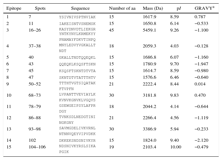
Based on the reactivity analysis of spots that were recognized by anti-ETX hyperimmune serum antibodies, 15 epitopic regions were identified in C. perfringens type D ETX (Table 1).
Reactive peptides in Clostridium perfringens type D ETX and each peptide's originating spot, sequence, number of amino acids (aa), molecular weight, isoelectric point (pI) and grand average of hydropathicity index (GRAVY).
| Epitope | Spots | Sequence | Number of aa | Mass (Da) | pI | GRAVYa |
|---|---|---|---|---|---|---|
| 1 | 7 | YSIVNIVSPTNVIAK | 15 | 1617.9 | 8.59 | 0.787 |
| 2 | 11 | IAKEISNTVSNEMSK | 15 | 1650.8 | 6.14 | −0.533 |
| 3 | 16–26 | KASYDNVDTLIEKGR YNTKYNYLKRMEKYY PNAMAYFDKVTINPQ | 45 | 5459.1 | 9.26 | −1.100 |
| 4 | 37–38 | MNYLEDVYVGKALLT NDT | 18 | 2059.3 | 4.03 | −0.128 |
| 5 | 40 | GKALLTNDTQQEQKL | 15 | 1686.8 | 6.07 | −1.160 |
| 6 | 43 | QQEQKLKSQSFTSKN | 15 | 1780.9 | 9.70 | −1.947 |
| 7 | 45 | KSQSFTSKNTDTVTA | 15 | 1614.7 | 8.59 | −0.980 |
| 8 | 47 | SKNTDTVTATTTHTV | 15 | 1576.6 | 6.46 | −0.640 |
| 9 | 50–52 | TTTHTVGTSIQATAK FTVPFN | 21 | 2222.4 | 8.44 | 0.014 |
| 10 | 68–73 | LVPANTTVEVIAYLK KVNVKGNVKLVGQVS | 30 | 3181.8 | 9.83 | 0.470 |
| 11 | 78–79 | GSEWGEIPSYLAFPR DGY | 18 | 2044.2 | 4.14 | −0.644 |
| 12 | 86–88 | TVNKSDLNEDGTINI NGKGNY | 21 | 2266.4 | 4.56 | −1.119 |
| 13 | 93–98 | SAVMGDELIVKVRNL NTNNVQEYVIPVDKK | 30 | 3386.9 | 5.94 | −0.233 |
| 14 | 102 | DKKEKSNDSNIVKYR | 15 | 1824.0 | 9.40 | −2.120 |
| 15 | 104–106 | NDSNIVKYRSLSIKA PGIK | 19 | 2103.4 | 10.00 | −0.479 |
Some of the probable determinants mapped were notable for their strong reactivity in immunochemical assays or due to existing data in the literature (described below) that corroborate the existence of these ETX epitopes. Epitope 3 (23LIEKGRYNTKYNYLKRMEKYY43) was the second most reactive epitope in immunochemical assays, which suggests that there are many antibodies against this epitope in the anti-ETX serum, which in turn, suggests that it is an immunodominant epitope. In addition, this epitope is located in domain I of C. perfringens type D ETX and is located in the amino-terminal region of the toxin (Fig. 2A). This region, which is mostly composed of a long alpha-helix, is believed to contain the toxin interaction site that binds to receptors present in target-cells.3,12 Thus, it is possible that neutralizing antibodies produced against ETX bind primarily to the amino-terminal region and prevent this toxin from adhering to cell receptors.
 Epitope 3. The yellow, green and red colors represent low, medium and high reactivity of regions, respectively. (B) Epitope 9 is shown in yellow; the determinant mapped by McClain and Cover15 is shown in black. (C) Epitope 9 is shown in yellow; the essential domain for ETX activity is shown in red; the overlapping region between this domain and epitope 9 is shown in orange. (D) Epitope 12 is shown in yellow; the negative electrostatic potential is shown in red; the positive electrostatic potential is shown in blue. (E) Epitope 15 is shown in yellow.")
View of the epitopes in the 3D structure of Clostridium perfringens type D ETX. (A) Epitope 3. The yellow, green and red colors represent low, medium and high reactivity of regions, respectively. (B) Epitope 9 is shown in yellow; the determinant mapped by McClain and Cover15 is shown in black. (C) Epitope 9 is shown in yellow; the essential domain for ETX activity is shown in red; the overlapping region between this domain and epitope 9 is shown in orange. (D) Epitope 12 is shown in yellow; the negative electrostatic potential is shown in red; the positive electrostatic potential is shown in blue. (E) Epitope 15 is shown in yellow.
Ivie and McClain13 introduced mutations in ETX by replacing seven accessible aromatic amino acids on its surface. Subsequently, they evaluated the ability of these mutants to interact with cells and to cause cell death. As a result, the authors observed that mutations in tyrosines Y42, Y43, Y49 and Y209 dramatically reduced protein binding to target-cells and that these residues are thus important for toxin cytotoxicity. Three tyrosines are present in the primary structure of epitope 3 (Y42, Y43, Y49), whereas the latter (Y209) is spatially close to the epitope (Fig. 2A), which therefore corroborates our results.
Determinant 9 (116TTTHTVGTSIQATAKFTVPFN136) contains the amino acid histidine (H) at position 119 of the primary ETX structure (Fig. 2B). According to Oyston et al.,14 when this residue is replaced by a proline, the toxin is inactivated. Thus, H119 and the region containing it are essential for the toxic activity of the protein, and anti-ETX antibodies against this epitope must neutralize this region.
Indeed, using two anti-ETX neutralizing monoclonal antibodies, McClain and Cover15 mapped an antigenic determinant close to epitope 9; this proximity occurs both linearly and spatially, as shown in Fig. 2B. According to the authors, when the monoclonal antibodies used bound to the identified determinant, they neutralized ETX by an allosteric mechanism, sterically preventing the loop containing H119 from exerting its function. However, according to these authors, the region between amino acids 119 and 158 is important for toxin activity, likely because it is the insertion region of the protein into the plasma membrane and participates in the formation of the transmembrane pore.
Knapp et al.16 also reported the importance of an ETX domain extending over the region from H119 to A149. Epitope 9 overlaps this domain at 18 amino acids, from H119 to N136 (Fig. 2C). According to the authors and corroborating the previously discussed findings, this region is involved in both toxin insertion into the membrane and cell pore formation, and mutations in some amino acids lead to changes in protein cytotoxicity and the characteristics of the formed pores.
Among the positive spots in the immunochemical assays, those corresponding to epitope 12 (224TVNKSDLNEDGTININGKGNY244) showed the highest reactivity, which suggests that many immunoglobulins against this antigenic domain are present in the anti-ETX sera. The GRAVY index of −1.119 of this epitope indicates high hydrophilicity, which in turn, predicts that this determinant is exceptionally accessible in ETX. This fact is corroborated by the position of epitope 12 in the 3D structure of the protein (Fig. 2D). Moreover, when calculating the electrostatic potential of the toxin, a large area with high potential is observed around a large portion of antigenic determinant 12 (Fig. 2D). Regions with electrostatic potential may form protein–protein or protein–ligand electrostatic interactions among themselves.17 In fact, these are major interactions that occur during the binding of an antibody to a protein antigen.18
Determinant 15 (282IVKYRSLSIKAPGIK296) is part of domain III in the carboxy-terminal portion of ETX (Fig. 2E), and this domain is believed to be involved in the interaction between the toxin chains during oligomerization to form the heptameric cell pore. In the native protein, the carboxy-terminal peptide seems to block the oligomerization of monomers and prevent the activation of the protoxin.19 Antibodies that bind to this region may competitively inhibit its cleavage by enzymes and then prevent the activation of the toxin. There are several examples described in the literature of antigens that have determinants in their carboxy-terminal portions,20–24 including C. botulinum toxin A4 and C. perfringens type A alpha toxin.2,25,26
The present study mapped 15 probable epitopes, with different immunodominance scales, consisting of part of the primary structure of C. perfringens type D ETX. Due to the strong reactivity in immunochemical assays and to existing knowledge in the literature, antigenic determinants 3, 9, 12 and 15 have a greater possibility of being immunodominant agents. Therefore, these determinants may be the basis for studies that aim to develop and formulate subunit vaccines against C. perfringens ETX.
Conflicts of interestThe authors declare that there are no conflicts of interest.
The authors would like to thank the Minas Gerais Research Support Foundation (Fundação de Amparo à Pesquisa do Estado de Minas Gerais – FAPEMIG), the Brazilian Federal Agency for the Support and Evaluation of Graduate Education (Coordenação de Aperfeiçoamento de Pessoal de Ensino Superior – CAPES) and the National Council for Scientific and Technological Development (Conselho Nacional de Desenvolvimento Científico e Tecnológico – CNPq).