


This communication reports the second known case of oral phaeohyphomycosis in a patient with squamocellular carcinoma of the lip. The patient, an 82-year-old black woman, a former smoker (for more than 30 years), suffering from an ulcerous vegetative lesion in the middle third of the lower lip for approximately 12 months. The result of the histopathological analysis indicated carcinoma, with well-differentiated keratinized squamous cells and the presence of septate mycelial filaments. In the direct mycological examination, thick and dematiaceous septate mycelial filaments were observed. After the resection surgery, the patient did not need to use an antifungal drug to treat the phaeohyphomycosis, and no follow-up radiotherapy was needed to treat the squamocellular carcinoma. We stress that the presence of the squamocellular lesion of the lip was a possible contributing factor to the infection.
Phaeohyphomycosis is a term used to denominate a set of cutaneous, subcutaneous and systemic infections caused by pigmented or dematiaceous fungi, producers of melanin. These fungi live in the soil and decomposing plant matter. They are the cause of sporadic cosmopolitan infections that afflict both healthy and immunosuppressed individuals. The main genera involved include Alternaria, Bipolaris, Cladophialophora, Cladosporium, Curvularia, Exophiala, Exserohilum, Phaeoacremonium, Phialophora and Wangiella.1,2
Generally the lesions originate from the inoculation point of fungal structures, through various traumas. They can remain local or spread through the adjacent tissues by hematogenic or lymphatic pathways. Whether or not infection will occur basically depends on three factors: resistance of the host, quantity of the inoculum and virulence of the fungus. With respect to virulence, it is believed that the melanin produced by the fungus improves the integrity of the cell walls and increases the total negative charge of the cells, protecting them against phagocytosis. Melanin can also protect the fungal cells against oxidative stress, extreme temperatures, iron depletion and microbial peptides.1,2
The diagnosis of phaeohyphomycosis is based on clinical observation, direct mycological examination using a potassium hydroxide (KOH) solution, isolation of the fungus in culture medium and histopathological analysis.1–3
This communication reports the second known case of oral phaeohyphomycosis in a patient with squamocellular carcinoma of the lip.
Case reportThe patientThe patient, an 82-year-old black woman, a former smoker (for more than 30 years), was attended by the head and neck outpatient service of Pernambuco Cancer Hospital, suffering from an ulcerous vegetative lesion in the middle third of the lower lip. The lesion measured approximately 2.5cm (with a “cauliflower” aspect) and had appeared about one year previously (Fig. 1A). For diagnosis of the etiology of the labial lesion, resection of the tissues for evaluation was indicated.
Diagnosis Ulcerative lesion of the lower lip. (B) View of the lower lip after surgical resection of the lesion.")
For the histological examination, a tissue fragment was stored in 10% formalin and sent to the hospital's pathology laboratory, where it was imbedded in paraffin and sliced into sections, which were stained with hematoxylin-eosin (HE) and periodic acid Schiff (PAS).
Another part of the clinical sample was sent for analysis at the medical mycology laboratory of the Center for Biological Sciences of Pernambuco Federal University, where direct mycological examination and culture analysis were carried out. A 20% aqueous solution of potassium hydroxide was used in the direct mycological examination for visualization of the fungal structures under an optical microscope. For culturing, the clinical sample was fragmented and inoculated in Petri dishes containing Sabouraud agar and brain heart infusion agar plus 50mg/L of chloramphenicol. The dishes were then maintained at 30°C and 37°C for over 30 days.
The result of the histopathological analysis indicated carcinoma, with well-differentiated keratinized squamous cells and the presence of septate mycelial filaments.
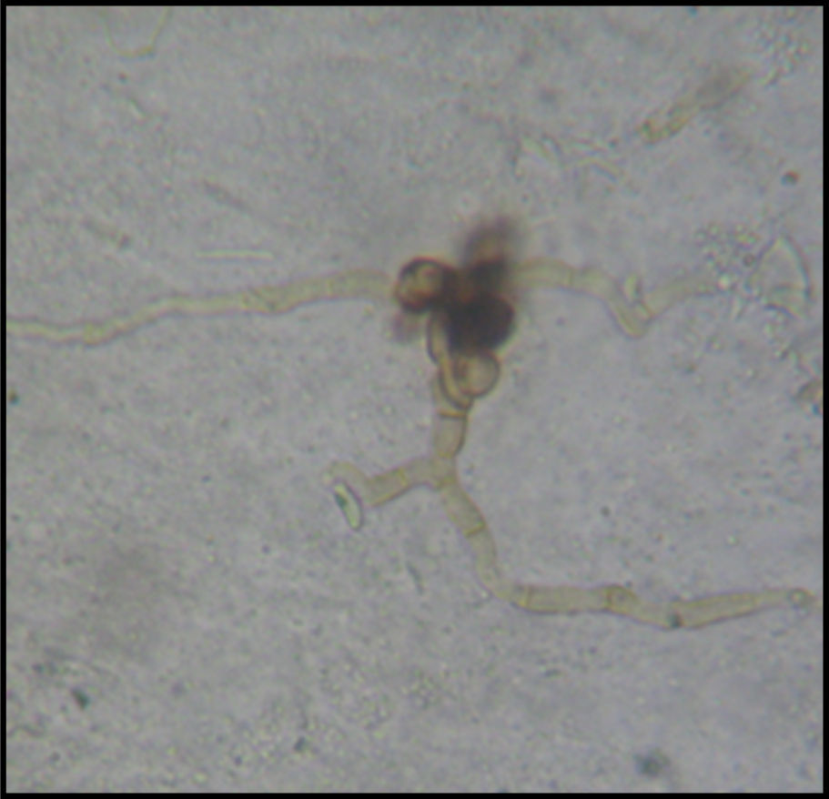
In the direct mycological examination, thick and dematiaceous septate mycelial filaments were observed (Fig. 2). There was no fungal growth in the culture media.

After the resection surgery, the patient did not need to use an antifungal drug to treat the phaeohyphomycosis, and no follow-up radiotherapy was needed to treat the squamocellular carcinoma (Fig. 1B).
DiscussionThe first case report of oral phaeohyphomycosis in the lip was published in 2007 by Cardoso and collaborators,4 in which a 57-year-old female patient presented a nodular lesion on the lower lip measuring 0.5cm in diameter. The patient reported she did not recall having suffered any type of injury, but stated she had used a herbal infusion to treat a renal infection. Besides drinking the herbal tea, she also had chewed the infused leaves. After the initial clinical evaluation, the team believed it was a case of pleomorphic adenoma, and surgical excision was proposed as treatment. However, the histopathological examination revealed the presence of septate hyphae and dematiaceous yeast-like structures, characterizing a case of oral phaeohyphomycosis. Through polymerase chain reaction (PCR), the authors confirmed the cause as being a fungus of the genus Alternaria. Despite this finding, treatment with antifungals was not necessary. The authors stressed that the type of clinical lesion afflicting the patient is very common and for this reason it is important to perform differential diagnosis.
Among the clinical manifestations of phaeohyphomycosis, superficial and subcutaneous infections are most common. Clinical cases of onychomycosis, tinea nigra, subcutaneous lesions, chromoblastomycosis, eumycetoma and keratitis have been reported in the medical literature. Besides superficial and subcutaneous infections, these fungi can also cause allergic reactions such as allergic fungal sinusitis and allergic bronchopulmonary mycosis. Among the most serious infections, dematiaceous fungi have been confirmed as etiological agents of pneumonia, brain abscess and disseminated disease. In these cases of high lethality and morbidity, the patients have presented some type of immunosuppression.1,2,5
Histopathalogical examination can help diagnose the ailment, by identifying inflammatory alterations and dematiaceous fungal elements.3 We found fungal structures in the histopathological examination. However, the direct mycological analysis produced more specific results (better visualization of the dematiaceous fungal structures). According to Cunha-Filho et al.,3 the diagnosis of phaeohyphomycosis should mainly be based on direct mycological examination, since the clinical appearance can be varied, the histopathology results unspecific and the culture negative (no fungal growth). In this case, it was not possible to isolate and identify the fungus because there was no growth in the culture medium. However, when analyzing the log of samples tested at the Medical Mycology Laboratory of UFPE, we found that the fungi involved in the phaeohyphomycosis cases were Cladosporium spp. and Curvularia spp. as agents of systemic infections, and Exophiala spp. and Phaeoacremonium spp. as agents of subcutaneous infections (data not shown).
There is no standard treatment for phaeohyphomycosis at present. In certain cases, complete surgical excision is sufficient for cure, with no need to administer antifungal drugs. In cases where antifungal therapy is necessary, itraconazole, voriconazole and posaconazole are the drugs of choice, through oral administration.6
Based on the findings in the literature, we believe this is the second report of a case of oral phaeohyphomycosis in the lip. There are only two other published reports, one of which describes a fungal infection of the palate and the other of the jaw, without any involvement of the lip.6,7 We stress that the presence of the lesion, with well-differentiated keratinized squamous cells was a possible contributing factor to the infection. Some dematiaceous filamentous fungi are known to have the ability to degrade keratin.2 In light of this case report, it can be inferred that more careful investigation is necessary regarding the association between cancerous lesions of the oral cavity and fungal infections. Recent studies have indicated that microbes, including fungi, can contribute to carcinogenesis in the oral mucosa as well as at other body sites.5,8,9
Conflicts of interestNone. The authors alone are responsible for the content and writing of the manuscript.
This work was supported by the Conselho Nacional de Desenvolvimento Científico e Tecnológico (CNPq).