VC-024 - COMBINED ENDOSCOPIC AND THORACOSCOPIC APPROACH FOR ESOPHAGEAL ENUCLEATION OF A SUBMUCOSAL BENIGN TUMOR
1Hospital Universitari Germans Trias i Pujol, Badalona; 2National Cancer Center Hospital, Tokio.
Introduction: Large submucosal tumors originating from the thoracic esophagus have a high risk of malignancy and may need surgery to obtain a definitive diagnosis and treatment. The more common approach for these large tumors is esophagectomy, a highly invasive procedure. We present a vídeo of a minimally invasive hybrid approach for a large submucosal tumor located on the thoracic esophagus, consisting of a combined endoscopic and thoracoscopic enucleation. This technique, described by Dr. Daiko et al., was published in the World Journal of Surgical Oncology in 2015.
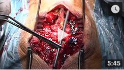
Case report: The patient of the vídeo is a 51 years old female diagnosed with a 40 mm submucosal tumor of the upper thoracic esophagus. Endoscopy with biopsies was performed but histologic results only showed inflammatory tissue. The study was completed with a barium swallow test, a neck and thorax CT scan and MRI showing a narrowed esophageal lumen around the tumor location. In order to have a definitive diagnosis of the submucosal tumor, surgery was indicated. A combined thoracoscopic and endoscopic approach was performed. The patient was in a left semi-prone position with the right arm raised cranially. Port position for the thoracoscopic part was the following: 5-mm-diameter ports at the second and forth intercostal spaces (ICS) on the mid-axillary line, 12-mm-diameter ports at the sixth ICS on the mid-axillary line and twelfth ICS on the posterior-axillary line. The first step of the thoracoscopy was a longitudinal incision at the mediastinal pleura, cranial to the azygos arch, for esophagus exposure. Next, the thoracic duct was individualized and the esophagus was mobilized to obtain a larger surgical field space. Simultaneously with thoracoscopy, the esophagoscopy started. A sodium hyaluronate solution with indigo carmine was endoscopically injected into the submucosal layer surrounding the tumor, highlighting its limits to perform a color-guided surgical incision. This solution also allowed to create space between the tumor and submucosal layer for the maximum esophageal mucosa layer-preservation. During the surgery, the tumor showed an important adhesion with the surrounding esophageal submucosa despite the injected solution in this area. Carefull dissection was performed, an accidental mucosal tear happened, which was repaired with a continuous suture. The rest of the enucleation procedure was carried out successfully. Surgery was performed within 90 minutes. The patient started gradual food intake on the third postoperative day after a non-pathologic endoscopy and barium swallow test, and was discharged on the seventh day with no evidence of complications. Histological diagnosis showed a schwannoma.
Discussion: The combined endoscopic and thoracoscopic approach for enucleation of large submucosal tumors on the thoracic esophagus offers accurate tumor delimitation and a better chance of preserving the esophageal mucosa than thoracoscopic enucleation alone, furthermore, it is safe and less invasive than esophagectomy. We encourage all esophageal surgeons to collaborate with endoscopists and take into consideration the described hybrid technique for patients diagnosed with large submucosal tumors of the thoracic esophagus, such as schwannoma, leiomyoma or GIST.


