array:24 [ "pii" => "S2173507722004264" "issn" => "21735077" "doi" => "10.1016/j.cireng.2022.12.003" "estado" => "S300" "fechaPublicacion" => "2023-11-01" "aid" => "2837" "copyright" => "AEC" "copyrightAnyo" => "2022" "documento" => "simple-article" "crossmark" => 1 "subdocumento" => "crp" "cita" => "Cir Esp. 2023;101:805-8" "abierto" => array:3 [ "ES" => false "ES2" => false "LATM" => false ] "gratuito" => false "lecturas" => array:1 [ "total" => 0 ] "Traduccion" => array:1 [ "es" => array:19 [ "pii" => "S0009739X22003554" "issn" => "0009739X" "doi" => "10.1016/j.ciresp.2022.10.024" "estado" => "S300" "fechaPublicacion" => "2023-11-01" "aid" => "2837" "copyright" => "AEC" "documento" => "simple-article" "crossmark" => 1 "subdocumento" => "crp" "cita" => "Cir Esp. 2023;101:805-8" "abierto" => array:3 [ "ES" => false "ES2" => false "LATM" => false ] "gratuito" => false "lecturas" => array:1 [ "total" => 0 ] "es" => array:11 [ "idiomaDefecto" => true "cabecera" => "<span class="elsevierStyleTextfn">Carta científica</span>" "titulo" => "Displasia de alto grado en el conducto cístico tras colecistectomía" "tienePdf" => "es" "tieneTextoCompleto" => "es" "paginas" => array:1 [ 0 => array:2 [ "paginaInicial" => "805" "paginaFinal" => "808" ] ] "titulosAlternativos" => array:1 [ "en" => array:1 [ "titulo" => "High-grade dysplasia in the cystic duct after cholecystectomy" ] ] "contieneTextoCompleto" => array:1 [ "es" => true ] "contienePdf" => array:1 [ "es" => true ] "resumenGrafico" => array:2 [ "original" => 0 "multimedia" => array:7 [ "identificador" => "fig0005" "etiqueta" => "Figura 1" "tipo" => "MULTIMEDIAFIGURA" "mostrarFloat" => true "mostrarDisplay" => false "figura" => array:1 [ 0 => array:4 [ "imagen" => "gr1.jpeg" "Alto" => 1132 "Ancho" => 2007 "Tamanyo" => 516737 ] ] "descripcion" => array:1 [ "es" => "<p id="spar0005" class="elsevierStyleSimplePara elsevierViewall">Displasia de alto grado epitelial en el borde de resección cístico de la pieza de colecistectomía mostrando pérdida de la polaridad nuclear y atipia intensa con irregularidad, agrandamiento e hipercromatismo nuclear. B y C) Vía biliar principal (flecha) en cortes coronales de RM en T1 (B) y T2 (C), sin objetivarse estenosis ni dilatación de la misma. D y E) Visión laparoscópica, objetivándose la vía biliar principal (flecha) y el muñón del conducto cístico (estrella) previa a su resección quirúrgica. F) Imagen tras la resección del remanente cístico, apreciándose vía biliar principal (flecha) con vena varicosa asociada (flecha punteada).</p>" ] ] ] "autores" => array:1 [ 0 => array:2 [ "autoresLista" => "Alberto García Picazo, Viviana Vega Novillo, Rosa González Martín, Alfredo Vivas López, Iago Justo Alonso" "autores" => array:5 [ 0 => array:2 [ "nombre" => "Alberto" "apellidos" => "García Picazo" ] 1 => array:2 [ "nombre" => "Viviana" "apellidos" => "Vega Novillo" ] 2 => array:2 [ "nombre" => "Rosa" "apellidos" => "González Martín" ] 3 => array:2 [ "nombre" => "Alfredo" "apellidos" => "Vivas López" ] 4 => array:2 [ "nombre" => "Iago Justo" "apellidos" => "Alonso" ] ] ] ] ] "idiomaDefecto" => "es" "Traduccion" => array:1 [ "en" => array:9 [ "pii" => "S2173507722004264" "doi" => "10.1016/j.cireng.2022.12.003" "estado" => "S300" "subdocumento" => "" "abierto" => array:3 [ "ES" => false "ES2" => false "LATM" => false ] "gratuito" => false "lecturas" => array:1 [ "total" => 0 ] "idiomaDefecto" => "en" "EPUB" => "https://multimedia.elsevier.es/PublicationsMultimediaV1/item/epub/S2173507722004264?idApp=UINPBA00004N" ] ] "EPUB" => "https://multimedia.elsevier.es/PublicationsMultimediaV1/item/epub/S0009739X22003554?idApp=UINPBA00004N" "url" => "/0009739X/0000010100000011/v1_202311020535/S0009739X22003554/v1_202311020535/es/main.assets" ] ] "itemSiguiente" => array:18 [ "pii" => "S2173507723000169" "issn" => "21735077" "doi" => "10.1016/j.cireng.2023.01.004" "estado" => "S300" "fechaPublicacion" => "2023-11-01" "aid" => "2839" "documento" => "simple-article" "crossmark" => 1 "subdocumento" => "crp" "cita" => "Cir Esp. 2023;101:808-11" "abierto" => array:3 [ "ES" => false "ES2" => false "LATM" => false ] "gratuito" => false "lecturas" => array:1 [ "total" => 0 ] "en" => array:11 [ "idiomaDefecto" => true "cabecera" => "<span class="elsevierStyleTextfn">Scientific letter</span>" "titulo" => "Sump syndrome as a late complication of side-to-side hepaticojejunostomy due to biliary iatrogenesis" "tienePdf" => "en" "tieneTextoCompleto" => "en" "paginas" => array:1 [ 0 => array:2 [ "paginaInicial" => "808" "paginaFinal" => "811" ] ] "titulosAlternativos" => array:1 [ "es" => array:1 [ "titulo" => "El síndrome de sumidero como complicación tardía de la hepaticoyeyunostomía latero-lateral por yatrogenia biliar" ] ] "contieneTextoCompleto" => array:1 [ "en" => true ] "contienePdf" => array:1 [ "en" => true ] "resumenGrafico" => array:2 [ "original" => 0 "multimedia" => array:8 [ "identificador" => "fig0005" "etiqueta" => "Fig. 1" "tipo" => "MULTIMEDIAFIGURA" "mostrarFloat" => true "mostrarDisplay" => false "figura" => array:1 [ 0 => array:4 [ "imagen" => "gr1.jpeg" "Alto" => 2158 "Ancho" => 2007 "Tamanyo" => 251824 ] ] "detalles" => array:1 [ 0 => array:3 [ "identificador" => "at0005" "detalle" => "Fig. " "rol" => "short" ] ] "descripcion" => array:1 [ "en" => "<p id="spar0005" class="elsevierStyleSimplePara elsevierViewall">(a) Sump syndrome in side-to-side hepaticojejunostomy due to accumulation of sludge and calculi in the common bile duct below the hepaticojejunal anastomosis. (b) Magnetic resonance cholangiopancreatography in the coronal plane showing dilation of the bile duct due to the presence of numerous lithiases (thin arrows) occupying the proximal extrahepatic and intrahepatic bile duct, in a patient with side-to-side choledochoduodenostomy (thick arrow). (c) Roux-en-Y end-to-side hepaticojejunostomy.</p>" ] ] ] "autores" => array:1 [ 0 => array:2 [ "autoresLista" => "Clara Fernández Fernández, Iago Justo Alonso, Andrea De Cimaa Fernández, Alberto Marcacuzco Quinto, Carlos Jiménez Romero" "autores" => array:5 [ 0 => array:2 [ "nombre" => "Clara" "apellidos" => "Fernández Fernández" ] 1 => array:2 [ "nombre" => "Iago" "apellidos" => "Justo Alonso" ] 2 => array:2 [ "nombre" => "Andrea" "apellidos" => "De Cimaa Fernández" ] 3 => array:2 [ "nombre" => "Alberto" "apellidos" => "Marcacuzco Quinto" ] 4 => array:2 [ "nombre" => "Carlos" "apellidos" => "Jiménez Romero" ] ] ] ] ] "idiomaDefecto" => "en" "Traduccion" => array:1 [ "es" => array:9 [ "pii" => "S0009739X22003578" "doi" => "10.1016/j.ciresp.2022.10.026" "estado" => "S300" "subdocumento" => "" "abierto" => array:3 [ "ES" => false "ES2" => false "LATM" => false ] "gratuito" => false "lecturas" => array:1 [ "total" => 0 ] "idiomaDefecto" => "es" "EPUB" => "https://multimedia.elsevier.es/PublicationsMultimediaV1/item/epub/S0009739X22003578?idApp=UINPBA00004N" ] ] "EPUB" => "https://multimedia.elsevier.es/PublicationsMultimediaV1/item/epub/S2173507723000169?idApp=UINPBA00004N" "url" => "/21735077/0000010100000011/v1_202311200506/S2173507723000169/v1_202311200506/en/main.assets" ] "itemAnterior" => array:19 [ "pii" => "S2173507722004276" "issn" => "21735077" "doi" => "10.1016/j.cireng.2022.12.004" "estado" => "S300" "fechaPublicacion" => "2023-11-01" "aid" => "2834" "copyright" => "AEC" "documento" => "simple-article" "crossmark" => 1 "subdocumento" => "crp" "cita" => "Cir Esp. 2023;101:802-5" "abierto" => array:3 [ "ES" => false "ES2" => false "LATM" => false ] "gratuito" => false "lecturas" => array:1 [ "total" => 0 ] "en" => array:10 [ "idiomaDefecto" => true "titulo" => "Spontaneous splenic rupture with massive bleeding during Pringle maneuver in laparoscopic liver resection" "tienePdf" => "en" "tieneTextoCompleto" => "en" "paginas" => array:1 [ 0 => array:2 [ "paginaInicial" => "802" "paginaFinal" => "805" ] ] "titulosAlternativos" => array:1 [ "es" => array:1 [ "titulo" => "Rotura esplénica espontánea con hemorragia masiva durante la maniobra de Pringle en cirugía hepática laparoscópica" ] ] "contieneTextoCompleto" => array:1 [ "en" => true ] "contienePdf" => array:1 [ "en" => true ] "resumenGrafico" => array:2 [ "original" => 0 "multimedia" => array:8 [ "identificador" => "fig0010" "etiqueta" => "Figure 2" "tipo" => "MULTIMEDIAFIGURA" "mostrarFloat" => true "mostrarDisplay" => false "figura" => array:1 [ 0 => array:4 [ "imagen" => "gr2.jpeg" "Alto" => 3682 "Ancho" => 2508 "Tamanyo" => 631369 ] ] "detalles" => array:1 [ 0 => array:3 [ "identificador" => "at0010" "detalle" => "Figure " "rol" => "short" ] ] "descripcion" => array:1 [ "en" => "<p id="spar0010" class="elsevierStyleSimplePara elsevierViewall">Physiopathology of portal hypertension.<a class="elsevierStyleCrossRef" href="#bib0030"><span class="elsevierStyleSup">6</span></a></p>" ] ] ] "autores" => array:1 [ 0 => array:2 [ "autoresLista" => "P. Camporro-González, N. Pujol-Cano, F.X. Molina-Romero, E. Palma-Zamora, J.M. Morón-Canis" "autores" => array:5 [ 0 => array:2 [ "nombre" => "P." "apellidos" => "Camporro-González" ] 1 => array:2 [ "nombre" => "N." "apellidos" => "Pujol-Cano" ] 2 => array:2 [ "nombre" => "F.X." "apellidos" => "Molina-Romero" ] 3 => array:2 [ "nombre" => "E." "apellidos" => "Palma-Zamora" ] 4 => array:2 [ "nombre" => "J.M." "apellidos" => "Morón-Canis" ] ] ] ] ] "idiomaDefecto" => "en" "Traduccion" => array:1 [ "es" => array:9 [ "pii" => "S0009739X22003529" "doi" => "10.1016/j.ciresp.2022.10.021" "estado" => "S300" "subdocumento" => "" "abierto" => array:3 [ "ES" => false "ES2" => false "LATM" => false ] "gratuito" => false "lecturas" => array:1 [ "total" => 0 ] "idiomaDefecto" => "es" "EPUB" => "https://multimedia.elsevier.es/PublicationsMultimediaV1/item/epub/S0009739X22003529?idApp=UINPBA00004N" ] ] "EPUB" => "https://multimedia.elsevier.es/PublicationsMultimediaV1/item/epub/S2173507722004276?idApp=UINPBA00004N" "url" => "/21735077/0000010100000011/v1_202311200506/S2173507722004276/v1_202311200506/en/main.assets" ] "en" => array:15 [ "idiomaDefecto" => true "cabecera" => "<span class="elsevierStyleTextfn">Scientific letter</span>" "titulo" => "High-grade dysplasia in the cystic duct after cholecystectomy" "tieneTextoCompleto" => true "paginas" => array:1 [ 0 => array:2 [ "paginaInicial" => "805" "paginaFinal" => "808" ] ] "autores" => array:1 [ 0 => array:4 [ "autoresLista" => "Alberto García Picazo, Viviana Vega Novillo, Rosa González Martín, Alfredo Vivas López, Iago Justo Alonso" "autores" => array:5 [ 0 => array:4 [ "nombre" => "Alberto García" "apellidos" => "Picazo" "email" => array:2 [ 0 => "agpicazo@salud.madrid.org" 1 => "alberto.gpicazo@gmail.com" ] "referencia" => array:1 [ 0 => array:2 [ "etiqueta" => "<span class="elsevierStyleSup">*</span>" "identificador" => "cor0005" ] ] ] 1 => array:2 [ "nombre" => "Viviana Vega" "apellidos" => "Novillo" ] 2 => array:2 [ "nombre" => "Rosa González" "apellidos" => "Martín" ] 3 => array:2 [ "nombre" => "Alfredo Vivas" "apellidos" => "López" ] 4 => array:2 [ "nombre" => "Iago Justo" "apellidos" => "Alonso" ] ] "afiliaciones" => array:1 [ 0 => array:2 [ "entidad" => "Servicio de Cirugía General, del Aparato Digestivo y de Trasplantes de Órganos Abdominales, Universidad Complutense de Madrid, Madrid, Spain" "identificador" => "aff0005" ] ] "correspondencia" => array:1 [ 0 => array:3 [ "identificador" => "cor0005" "etiqueta" => "⁎" "correspondencia" => "<span class="elsevierStyleItalic">Corresponding author</span>." ] ] ] ] "titulosAlternativos" => array:1 [ "es" => array:1 [ "titulo" => "Displasia de alto grado en el conducto cístico tras colecistectomía" ] ] "resumenGrafico" => array:2 [ "original" => 0 "multimedia" => array:8 [ "identificador" => "fig0005" "etiqueta" => "Fig. 1" "tipo" => "MULTIMEDIAFIGURA" "mostrarFloat" => true "mostrarDisplay" => false "figura" => array:1 [ 0 => array:4 [ "imagen" => "gr1.jpeg" "Alto" => 1132 "Ancho" => 2007 "Tamanyo" => 506409 ] ] "detalles" => array:1 [ 0 => array:3 [ "identificador" => "at0005" "detalle" => "Fig. " "rol" => "short" ] ] "descripcion" => array:1 [ "en" => "<p id="spar0005" class="elsevierStyleSimplePara elsevierViewall">High-grade epithelial dysplasia in the cystic duct resection margin of the cholecystectomy specimen, showing loss of nuclear polarity and intense atypia with nuclear hyperchromatism, irregularity and enlargement; B and C) Main bile duct (Arrow) in coronal MRI slices in T1 (B) and T2 (C), with no observed stenosis or dilatation; D and E) Laparoscopic view, showing the main bile duct (arrow) and the stump of the cystic duct (Star) prior to its surgical resection; F) Image after resection of the cystic remnant, showing the main bile duct (arrow) with associated varicose vein (dotted arrow).</p>" ] ] ] "textoCompleto" => "<span class="elsevierStyleSections"><p id="par0005" class="elsevierStylePara elsevierViewall">Laparoscopic cholecystectomy is one of the most frequently performed surgeries. High-grade dysplasia (HGD) in the cystic duct resection margin is uncommon after cholecystectomy for cholelithiasis (<0.1%) or acute cholecystitis (1%), and it is considered a precursor of gallbladder and bile duct cancer.<a class="elsevierStyleCrossRefs" href="#bib0005"><span class="elsevierStyleSup">1,2</span></a> Therefore, when there is a finding of dysplasia, it is necessary to rule out concomitant tumor pathology, mainly multifocal biliary tumors (biliary intraductal papillary neoplasm, gallbladder adenocarcinoma and cholangiocarcinoma).<a class="elsevierStyleCrossRef" href="#bib0010"><span class="elsevierStyleSup">2</span></a> However, when faced with an isolated HGD with no clear signs of invasion, there is no therapeutic consensus.<a class="elsevierStyleCrossRef" href="#bib0015"><span class="elsevierStyleSup">3</span></a></p><p id="par0010" class="elsevierStylePara elsevierViewall">
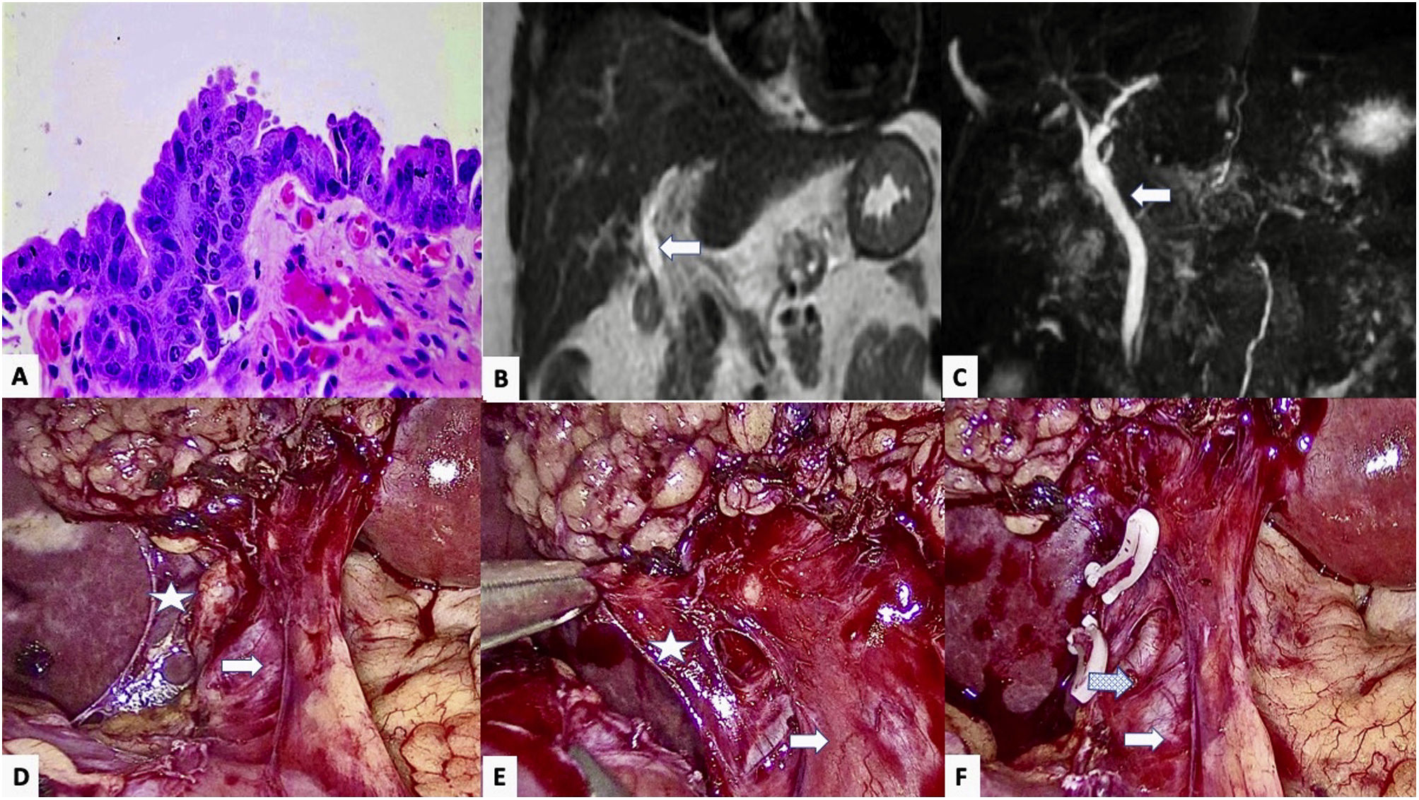
We present a 44-year-old woman with a medical history of multiple sclerosis, bone marrow transplant for acute myeloid leukemia, and cholelithiasis with various episodes of biliary colic over the course of a year, for which she underwent elective laparoscopic cholecystectomy (performed without incident). The histopathological study revealed a 2-mm high-grade dysplasia (high-grade biliary intraepithelial neoplasm) in the resection margin of the cystic duct, yet no involvement of the gallbladder (<a class="elsevierStyleCrossRef" href="#fig0005">Fig. 1</a>A). On physical examination, the patient presented no abdominal pain or jaundice, nor did she report weight loss. The blood work-up was normal, showing no changes in liver enzymes or bilirubin, and tumor marker levels were normal (CEA 3.09 ng/mL, Ca 19.9 18.80 U/mL). An MR cholangiopancreatography (MRCP) of the liver detected no space-occupying lesion (SOL), pathological lymphadenopathies, or dilation of the bile duct (<a class="elsevierStyleCrossRef" href="#fig0005">Fig. 1</a>B and C).</p><elsevierMultimedia ident="fig0005"></elsevierMultimedia><p id="par0015" class="elsevierStylePara elsevierViewall">Based on the histology report and the absence of distant disease, the multidisciplinary committee ruled out any further diagnostic studies (CT, ERCP) and proposed a surgical intervention for the re-resection of the cystic margin. The procedure was laparoscopic and entailed dissection and division of the cystic remnant, proximal to the bile duct (<a class="elsevierStyleCrossRef" href="#fig0005">Fig. 1</a>D and F). The intraoperative study of the resection margin (1 × 0.6 × 0.1 cm) as well as the re-resection of the cystic gland showed no residual dysplasia or malignancy. The postoperative period was uneventful. One month after surgery, the follow-up (MRCP) revealed nothing of interest.</p><p id="par0020" class="elsevierStylePara elsevierViewall">The appearance of HGD in the cystic margin after cholecystectomy is a rare finding (0.2%–1%).<a class="elsevierStyleCrossRefs" href="#bib0015"><span class="elsevierStyleSup">3,4</span></a> Its importance lies in the potential progression to carcinoma (cholangiocarcinoma in the bile duct or gallbladder adenocarcinoma), as it is a common precursor (69% of cases) and both have an ominous prognosis with invasion.<a class="elsevierStyleCrossRefs" href="#bib0015"><span class="elsevierStyleSup">3,4</span></a> If the diagnosis of malignancy is confirmed, surgical treatment must be aggressive and include: bile duct resection; hepatectomy of segments IVb and V; and lymph node dissection of the portal, perihilar, and gastrohepatic ligament nodes.<a class="elsevierStyleCrossRef" href="#bib0005"><span class="elsevierStyleSup">1</span></a> Some groups have described a correlation of HGD with extrahepatic cholangiocarcinoma (13%–20%) and also report that that the main prognostic factor for survival was surgical margin involvement.<a class="elsevierStyleCrossRefs" href="#bib0020"><span class="elsevierStyleSup">4–6</span></a> Due to the multifocal nature of tumors in this location, exploration of the bile duct is mandatory, requiring resection in many cases (<a class="elsevierStyleCrossRef" href="#tbl0005">Table 1</a>).<a class="elsevierStyleCrossRef" href="#bib0035"><span class="elsevierStyleSup">7</span></a></p><elsevierMultimedia ident="tbl0005"></elsevierMultimedia><p id="par0025" class="elsevierStylePara elsevierViewall">However, the presence of HGD when there is no suspected malignancy is quite rare (0.05%).<a class="elsevierStyleCrossRefs" href="#bib0040"><span class="elsevierStyleSup">8,9</span></a> In these patients, the main objective, as highlighted by the guidelines of the American Hepato-Pancreato-Biliary Association (AHPBA), is to rule out adjacent tumor pathology,<a class="elsevierStyleCrossRef" href="#bib0050"><span class="elsevierStyleSup">10</span></a> initially based on intraoperative findings of the previous cholecystectomy, presence of lymphadenopathies, and dilation of the bile duct, gallbladder or biliary tumor. Secondly, after the histological confirmation of HGD, an extension study is necessary with abdominal CT and MRCP. Tumor markers (CEA, CA 19.9) could help guide the diagnosis. As previously mentioned, this type of neoplasm tends to be multifocal, so a diagnostic ERCP could be appropriate.</p><p id="par0030" class="elsevierStylePara elsevierViewall">Preoperative diagnostic tests frequently do not provide relevant information. Thus, the question that remains is whether radical surgery is necessary or whether cystic resection and close surveillance would suffice in the absence of diagnosis or evidence of malignancy. In this context, the role of multidisciplinary committees becomes essential. Until now, radical surgery with excision of the bile duct has been considered the appropriate therapy. However, the most recent published papers advocate a conservative approach with close monitoring, either with or without surgical treatment.<a class="elsevierStyleCrossRefs" href="#bib0005"><span class="elsevierStyleSup">1,4</span></a> Due to its importance, this decision must always be made with the patient, most whom opt for surgical treatment, as described in other series.<a class="elsevierStyleCrossRef" href="#bib0020"><span class="elsevierStyleSup">4</span></a> The diagnosis is histological, based on the detection of cells with HGD, suggesting that the disease-free resection margin should be at least 5 mm, in prognostic terms.</p><p id="par0035" class="elsevierStylePara elsevierViewall">In our case, we decided to reoperate with minimally invasive surgery, resecting the cystic remnant and confirming the absence of margin involvement with an intraoperative sample. This latter technique is not mandatory due to its low diagnostic yield (fibrosis, inflammatory changes), but it helps determine the prognosis and extension of surgery.<a class="elsevierStyleCrossRef" href="#bib0015"><span class="elsevierStyleSup">3</span></a> Regarding postoperative follow-up, the data in the literature are ambiguous. Nevertheless, a follow-up of at least 5 years is recommended, similar to biliary carcinoma, with tumor markers (CEA, CA 19.9) and abdominal ultrasound/CT scan every 6 months for 2 years, and annually thereafter.</p><p id="par0040" class="elsevierStylePara elsevierViewall">In conclusion, HGD of the cystic remnant with no evidence of malignancy after cholecystectomy is a rare pathology. However, it is necessary to rule out its association with biliary tumor pathology, and the treatment of choice includes cystic resection and close postoperative follow-up.</p><span id="sec0005" class="elsevierStyleSection elsevierViewall"><span class="elsevierStyleSectionTitle" id="sect0005">Conflicts of interest</span><p id="par0045" class="elsevierStylePara elsevierViewall">None of the authors have any conflicts of interests.</p></span></span>" "textoCompletoSecciones" => array:1 [ "secciones" => array:3 [ 0 => array:2 [ "identificador" => "sec0005" "titulo" => "Conflicts of interest" ] 1 => array:2 [ "identificador" => "xack704915" "titulo" => "Acknowledgements" ] 2 => array:1 [ "titulo" => "References" ] ] ] "pdfFichero" => "main.pdf" "tienePdf" => true "multimedia" => array:2 [ 0 => array:8 [ "identificador" => "fig0005" "etiqueta" => "Fig. 1" "tipo" => "MULTIMEDIAFIGURA" "mostrarFloat" => true "mostrarDisplay" => false "figura" => array:1 [ 0 => array:4 [ "imagen" => "gr1.jpeg" "Alto" => 1132 "Ancho" => 2007 "Tamanyo" => 506409 ] ] "detalles" => array:1 [ 0 => array:3 [ "identificador" => "at0005" "detalle" => "Fig. " "rol" => "short" ] ] "descripcion" => array:1 [ "en" => "<p id="spar0005" class="elsevierStyleSimplePara elsevierViewall">High-grade epithelial dysplasia in the cystic duct resection margin of the cholecystectomy specimen, showing loss of nuclear polarity and intense atypia with nuclear hyperchromatism, irregularity and enlargement; B and C) Main bile duct (Arrow) in coronal MRI slices in T1 (B) and T2 (C), with no observed stenosis or dilatation; D and E) Laparoscopic view, showing the main bile duct (arrow) and the stump of the cystic duct (Star) prior to its surgical resection; F) Image after resection of the cystic remnant, showing the main bile duct (arrow) with associated varicose vein (dotted arrow).</p>" ] ] 1 => array:8 [ "identificador" => "tbl0005" "etiqueta" => "Table 1" "tipo" => "MULTIMEDIATABLA" "mostrarFloat" => true "mostrarDisplay" => false "detalles" => array:1 [ 0 => array:3 [ "identificador" => "at0010" "detalle" => "Table " "rol" => "short" ] ] "tabla" => array:1 [ "tablatextoimagen" => array:1 [ 0 => array:2 [ "tabla" => array:1 [ 0 => """ <table border="0" frame="\n \t\t\t\t\tvoid\n \t\t\t\t" class=""><thead title="thead"><tr title="table-row"><th class="td" title="\n \t\t\t\t\ttable-head\n \t\t\t\t " align="left" valign="\n \t\t\t\t\ttop\n \t\t\t\t" scope="col" style="border-bottom: 2px solid black">Cases \t\t\t\t\t\t\n \t\t\t\t\t\t</th><th class="td" title="\n \t\t\t\t\ttable-head\n \t\t\t\t " align="center" valign="\n \t\t\t\t\ttop\n \t\t\t\t" scope="col" style="border-bottom: 2px solid black">Age (years) \t\t\t\t\t\t\n \t\t\t\t\t\t</th><th class="td" title="\n \t\t\t\t\ttable-head\n \t\t\t\t " align="center" valign="\n \t\t\t\t\ttop\n \t\t\t\t" scope="col" style="border-bottom: 2px solid black">Sex \t\t\t\t\t\t\n \t\t\t\t\t\t</th><th class="td" title="\n \t\t\t\t\ttable-head\n \t\t\t\t " align="center" valign="\n \t\t\t\t\ttop\n \t\t\t\t" scope="col" style="border-bottom: 2px solid black">Preoperative image \t\t\t\t\t\t\n \t\t\t\t\t\t</th><th class="td" title="\n \t\t\t\t\ttable-head\n \t\t\t\t " align="center" valign="\n \t\t\t\t\ttop\n \t\t\t\t" scope="col" style="border-bottom: 2px solid black">Surgery \t\t\t\t\t\t\n \t\t\t\t\t\t</th><th class="td" title="\n \t\t\t\t\ttable-head\n \t\t\t\t " align="center" valign="\n \t\t\t\t\ttop\n \t\t\t\t" scope="col" style="border-bottom: 2px solid black">Histology \t\t\t\t\t\t\n \t\t\t\t\t\t</th><th class="td" title="\n \t\t\t\t\ttable-head\n \t\t\t\t " align="center" valign="\n \t\t\t\t\ttop\n \t\t\t\t" scope="col" style="border-bottom: 2px solid black">Survival \t\t\t\t\t\t\n \t\t\t\t\t\t</th></tr></thead><tbody title="tbody"><tr title="table-row"><td class="td-with-role" title="\n \t\t\t\t\ttable-entry\n \t\t\t\t ; entry_with_role_rowhead " align="left" valign="\n \t\t\t\t\ttop\n \t\t\t\t">Bickenbach KA et all, 2011. (N = 5) \t\t\t\t\t\t\n \t\t\t\t</td><td class="td" title="\n \t\t\t\t\ttable-entry\n \t\t\t\t " align="left" valign="\n \t\t\t\t\ttop\n \t\t\t\t">65.2 (57−77) \t\t\t\t\t\t\n \t\t\t\t</td><td class="td" title="\n \t\t\t\t\ttable-entry\n \t\t\t\t " align="left" valign="\n \t\t\t\t\ttop\n \t\t\t\t">Males (3:5) \t\t\t\t\t\t\n \t\t\t\t</td><td class="td" title="\n \t\t\t\t\ttable-entry\n \t\t\t\t " align="left" valign="\n \t\t\t\t\ttop\n \t\t\t\t">80% MRI40% CT + MRI20% ERCP + MRI \t\t\t\t\t\t\n \t\t\t\t</td><td class="td" title="\n \t\t\t\t\ttable-entry\n \t\t\t\t " align="left" valign="\n \t\t\t\t\ttop\n \t\t\t\t">Resection of cystic+/− (80%) lymphadenectomy +/− (40%) segmentectomy IV-V \t\t\t\t\t\t\n \t\t\t\t</td><td class="td" title="\n \t\t\t\t\ttable-entry\n \t\t\t\t " align="left" valign="\n \t\t\t\t\ttop\n \t\t\t\t">80% no dysplasia20% cholangiocarcinoma (T2N1) \t\t\t\t\t\t\n \t\t\t\t</td><td class="td" title="\n \t\t\t\t\ttable-entry\n \t\t\t\t " align="left" valign="\n \t\t\t\t\ttop\n \t\t\t\t">80% disease-free20% death \t\t\t\t\t\t\n \t\t\t\t</td></tr><tr title="table-row"><td class="td-with-role" title="\n \t\t\t\t\ttable-entry\n \t\t\t\t ; entry_with_role_rowhead " align="left" valign="\n \t\t\t\t\ttop\n \t\t\t\t">Sunkel-Laing B et al, 2014. (N = 1) \t\t\t\t\t\t\n \t\t\t\t</td><td class="td" title="\n \t\t\t\t\ttable-entry\n \t\t\t\t " align="left" valign="\n \t\t\t\t\ttop\n \t\t\t\t">67 \t\t\t\t\t\t\n \t\t\t\t</td><td class="td" title="\n \t\t\t\t\ttable-entry\n \t\t\t\t " align="left" valign="\n \t\t\t\t\ttop\n \t\t\t\t">Male \t\t\t\t\t\t\n \t\t\t\t</td><td class="td" title="\n \t\t\t\t\ttable-entry\n \t\t\t\t " align="left" valign="\n \t\t\t\t\ttop\n \t\t\t\t">CT + ERCP \t\t\t\t\t\t\n \t\t\t\t</td><td class="td" title="\n \t\t\t\t\ttable-entry\n \t\t\t\t " align="left" valign="\n \t\t\t\t\ttop\n \t\t\t\t">Pancreaticoduodenectomy (Whipple) \t\t\t\t\t\t\n \t\t\t\t</td><td class="td" title="\n \t\t\t\t\ttable-entry\n \t\t\t\t " align="left" valign="\n \t\t\t\t\ttop\n \t\t\t\t">Cholangiocarcinoma \t\t\t\t\t\t\n \t\t\t\t</td><td class="td" title="\n \t\t\t\t\ttable-entry\n \t\t\t\t " align="left" valign="\n \t\t\t\t\ttop\n \t\t\t\t">Death \t\t\t\t\t\t\n \t\t\t\t</td></tr><tr title="table-row"><td class="td-with-role" title="\n \t\t\t\t\ttable-entry\n \t\t\t\t ; entry_with_role_rowhead " align="left" valign="\n \t\t\t\t\ttop\n \t\t\t\t">Moslim MA et al, 2017. (N = 1) \t\t\t\t\t\t\n \t\t\t\t</td><td class="td" title="\n \t\t\t\t\ttable-entry\n \t\t\t\t " align="left" valign="\n \t\t\t\t\ttop\n \t\t\t\t">36 \t\t\t\t\t\t\n \t\t\t\t</td><td class="td" title="\n \t\t\t\t\ttable-entry\n \t\t\t\t " align="left" valign="\n \t\t\t\t\ttop\n \t\t\t\t">Female \t\t\t\t\t\t\n \t\t\t\t</td><td class="td" title="\n \t\t\t\t\ttable-entry\n \t\t\t\t " align="left" valign="\n \t\t\t\t\ttop\n \t\t\t\t">MRI + ERCP + CT \t\t\t\t\t\t\n \t\t\t\t</td><td class="td" title="\n \t\t\t\t\ttable-entry\n \t\t\t\t " align="left" valign="\n \t\t\t\t\ttop\n \t\t\t\t">Cystic resection \t\t\t\t\t\t\n \t\t\t\t</td><td class="td" title="\n \t\t\t\t\ttable-entry\n \t\t\t\t " align="left" valign="\n \t\t\t\t\ttop\n \t\t\t\t">No dysplasia \t\t\t\t\t\t\n \t\t\t\t</td><td class="td" title="\n \t\t\t\t\ttable-entry\n \t\t\t\t " align="left" valign="\n \t\t\t\t\ttop\n \t\t\t\t">Disease-free \t\t\t\t\t\t\n \t\t\t\t</td></tr><tr title="table-row"><td class="td-with-role" title="\n \t\t\t\t\ttable-entry\n \t\t\t\t ; entry_with_role_rowhead " align="left" valign="\n \t\t\t\t\ttop\n \t\t\t\t">Saliba M et al, 2019. (N = 1) \t\t\t\t\t\t\n \t\t\t\t</td><td class="td" title="\n \t\t\t\t\ttable-entry\n \t\t\t\t " align="left" valign="\n \t\t\t\t\ttop\n \t\t\t\t">44 \t\t\t\t\t\t\n \t\t\t\t</td><td class="td" title="\n \t\t\t\t\ttable-entry\n \t\t\t\t " align="left" valign="\n \t\t\t\t\ttop\n \t\t\t\t">Male \t\t\t\t\t\t\n \t\t\t\t</td><td class="td" title="\n \t\t\t\t\ttable-entry\n \t\t\t\t " align="left" valign="\n \t\t\t\t\ttop\n \t\t\t\t">MRI \t\t\t\t\t\t\n \t\t\t\t</td><td class="td" title="\n \t\t\t\t\ttable-entry\n \t\t\t\t " align="left" valign="\n \t\t\t\t\ttop\n \t\t\t\t">Cystic resection \t\t\t\t\t\t\n \t\t\t\t</td><td class="td" title="\n \t\t\t\t\ttable-entry\n \t\t\t\t " align="left" valign="\n \t\t\t\t\ttop\n \t\t\t\t">No dysplasia \t\t\t\t\t\t\n \t\t\t\t</td><td class="td" title="\n \t\t\t\t\ttable-entry\n \t\t\t\t " align="left" valign="\n \t\t\t\t\ttop\n \t\t\t\t">Disease-free \t\t\t\t\t\t\n \t\t\t\t</td></tr></tbody></table> """ ] "imagenFichero" => array:1 [ 0 => "xTab3350450.png" ] ] ] ] "descripcion" => array:1 [ "en" => "<p id="spar0010" class="elsevierStyleSimplePara elsevierViewall">
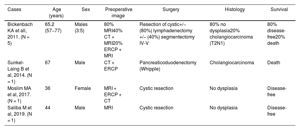
Distribution of the HGD cases described in the literature.</p>" ] ] ] "bibliografia" => array:2 [ "titulo" => "References" "seccion" => array:1 [ 0 => array:2 [ "identificador" => "bibs0005" "bibliografiaReferencia" => array:10 [ 0 => array:3 [ "identificador" => "bib0005" "etiqueta" => "1" "referencia" => array:1 [ 0 => array:2 [ "contribucion" => array:1 [ 0 => array:2 [ "titulo" => "Management of high-grade dysplasia of the cystic duct after cholecystectomy" "autores" => array:1 [ 0 => array:2 [ "etal" => false "autores" => array:3 [ 0 => "M.A. Moslim" 1 => "A. Tang" 2 => "G. Morris-Stiff" ] ] ] ] ] "host" => array:1 [ 0 => array:2 [ "doi" => "10.1136/bcr-2016-218994" "Revista" => array:2 [ "tituloSerie" => "BMJ Case Rep." "fecha" => "2017" ] ] ] ] ] ] 1 => array:3 [ "identificador" => "bib0010" "etiqueta" => "2" "referencia" => array:1 [ 0 => array:2 [ "contribucion" => array:1 [ 0 => array:2 [ "titulo" => "Predictive factors for incidental gallbladder dysplasia and carcinoma" "autores" => array:1 [ 0 => array:2 [ "etal" => false "autores" => array:6 [ 0 => "L. Solaini" 1 => "A. Sharma" 2 => "J. Watt" 3 => "S. Iosifidou" 4 => "J.A. Chin Aleong" 5 => "H.M. Kocher" ] ] ] ] ] "host" => array:1 [ 0 => array:2 [ "doi" => "10.1016/j.jss.2014.01.064" "Revista" => array:6 [ "tituloSerie" => "J Surg Res." "fecha" => "2014" "volumen" => "189" "paginaInicial" => "17" "paginaFinal" => "21" "link" => array:1 [ 0 => array:2 [ "url" => "https://www.ncbi.nlm.nih.gov/pubmed/24589178" "web" => "Medline" ] ] ] ] ] ] ] ] 2 => array:3 [ "identificador" => "bib0015" "etiqueta" => "3" "referencia" => array:1 [ 0 => array:2 [ "contribucion" => array:1 [ 0 => array:2 [ "titulo" => "Incidental high-grade displasia of the cystic duct margin: case report and literature review" "autores" => array:1 [ 0 => array:2 [ "etal" => false "autores" => array:2 [ 0 => "M. Saliba" 1 => "G. Zaatari" ] ] ] ] ] "host" => array:1 [ 0 => array:2 [ "doi" => "10.21037/cco.2019.07.04" "Revista" => array:6 [ "tituloSerie" => "Chin Clin Oncol." "fecha" => "2019" "volumen" => "8" "numero" => "4" "paginaInicial" => "34" "link" => array:1 [ 0 => array:2 [ "url" => "https://www.ncbi.nlm.nih.gov/pubmed/31431033" "web" => "Medline" ] ] ] ] ] ] ] ] 3 => array:3 [ "identificador" => "bib0020" "etiqueta" => "4" "referencia" => array:1 [ 0 => array:2 [ "contribucion" => array:1 [ 0 => array:2 [ "titulo" => "High-grade dysplasia of the cystic duct margin in the absence of malignancy after cholecystectomy" "autores" => array:1 [ 0 => array:2 [ "etal" => true "autores" => array:6 [ 0 => "K.A. Bickenbach" 1 => "J. Shia" 2 => "D.S. Klimstra" 3 => "R.P. DeMatteo" 4 => "Y. Fong" 5 => "T.P. Kingham" ] ] ] ] ] "host" => array:1 [ 0 => array:1 [ "Revista" => array:5 [ "tituloSerie" => "HPB." "fecha" => "2011" "volumen" => "13" "paginaInicial" => "865" "paginaFinal" => "868" ] ] ] ] ] ] 4 => array:3 [ "identificador" => "bib0025" "etiqueta" => "5" "referencia" => array:1 [ 0 => array:2 [ "contribucion" => array:1 [ 0 => array:2 [ "titulo" => "Clinical value of additional resection of a margin-positive proximal bile duct in hilar cholangiocarcinoma" "autores" => array:1 [ 0 => array:2 [ "etal" => false "autores" => array:6 [ 0 => "Y. Shingu" 1 => "T. Ebata" 2 => "H. Nishio" 3 => "T. Igami" 4 => "Y. Shimoyama" 5 => "M. Nagino" ] ] ] ] ] "host" => array:1 [ 0 => array:2 [ "doi" => "10.1016/j.surg.2009.06.030" "Revista" => array:6 [ "tituloSerie" => "Surgery." "fecha" => "2010" "volumen" => "147" "paginaInicial" => "49" "paginaFinal" => "56" "link" => array:1 [ 0 => array:2 [ "url" => "https://www.ncbi.nlm.nih.gov/pubmed/19767048" "web" => "Medline" ] ] ] ] ] ] ] ] 5 => array:3 [ "identificador" => "bib0030" "etiqueta" => "6" "referencia" => array:1 [ 0 => array:2 [ "contribucion" => array:1 [ 0 => array:2 [ "titulo" => "Impact of ductal resection margin status on longterm survival in patients undergoing resection for extrahepatic cholangiocarcinoma" "autores" => array:1 [ 0 => array:2 [ "etal" => false "autores" => array:5 [ 0 => "T. Wakai" 1 => "Y. Shirai" 2 => "T. Moroda" 3 => "N. Yokoyama" 4 => "K. Hatakeyama" ] ] ] ] ] "host" => array:1 [ 0 => array:2 [ "doi" => "10.1002/cncr.20906" "Revista" => array:6 [ "tituloSerie" => "Cancer." "fecha" => "2005" "volumen" => "103" "paginaInicial" => "1210" "paginaFinal" => "1216" "link" => array:1 [ 0 => array:2 [ "url" => "https://www.ncbi.nlm.nih.gov/pubmed/15685618" "web" => "Medline" ] ] ] ] ] ] ] ] 6 => array:3 [ "identificador" => "bib0035" "etiqueta" => "7" "referencia" => array:1 [ 0 => array:2 [ "contribucion" => array:1 [ 0 => array:2 [ "titulo" => "Repeated negative biopsies in isolated high-grade cystic duct dysplasia with progression to adenocarcinoma" "autores" => array:1 [ 0 => array:2 [ "etal" => false "autores" => array:5 [ 0 => "B. Sunkel-Laing" 1 => "A. Kalam" 2 => "I. Renfrew" 3 => "L. Mears" 4 => "H.M. Kocher" ] ] ] ] ] "host" => array:1 [ 0 => array:2 [ "doi" => "10.1159/000368114" "Revista" => array:6 [ "tituloSerie" => "Case Rep Gastroenterol." "fecha" => "2014" "volumen" => "8" "paginaInicial" => "304" "paginaFinal" => "309" "link" => array:1 [ 0 => array:2 [ "url" => "https://www.ncbi.nlm.nih.gov/pubmed/25473388" "web" => "Medline" ] ] ] ] ] ] ] ] 7 => array:3 [ "identificador" => "bib0040" "etiqueta" => "8" "referencia" => array:1 [ 0 => array:2 [ "contribucion" => array:1 [ 0 => array:2 [ "titulo" => "Distribution of dysplasia and cancer in the gallbladder: an analysis from a high cancer-risk population" "autores" => array:1 [ 0 => array:2 [ "etal" => true "autores" => array:6 [ 0 => "J. Koshiol" 1 => "E. Bellolio" 2 => "C. Vivallo" 3 => "P. Cook" 4 => "J.C. Roa" 5 => "E.E. McGee" ] ] ] ] ] "host" => array:1 [ 0 => array:2 [ "doi" => "10.1016/j.humpath.2018.07.015" "Revista" => array:6 [ "tituloSerie" => "Hum Pathol." "fecha" => "2018" "volumen" => "82" "paginaInicial" => "87" "paginaFinal" => "94" "link" => array:1 [ 0 => array:2 [ "url" => "https://www.ncbi.nlm.nih.gov/pubmed/30036595" "web" => "Medline" ] ] ] ] ] ] ] ] 8 => array:3 [ "identificador" => "bib0045" "etiqueta" => "9" "referencia" => array:1 [ 0 => array:2 [ "contribucion" => array:1 [ 0 => array:2 [ "titulo" => "Submitting the entire gallbladder in cases of dysplasia is not justified" "autores" => array:1 [ 0 => array:2 [ "etal" => false "autores" => array:2 [ 0 => "A.A. Renshaw" 1 => "E.W. Gould" ] ] ] ] ] "host" => array:1 [ 0 => array:2 [ "doi" => "10.1309/AJCPB0ZTXXIF6MOF" "Revista" => array:6 [ "tituloSerie" => "Am J Clin Pathol." "fecha" => "2012" "volumen" => "138" "paginaInicial" => "374" "paginaFinal" => "376" "link" => array:1 [ 0 => array:2 [ "url" => "https://www.ncbi.nlm.nih.gov/pubmed/22912353" "web" => "Medline" ] ] ] ] ] ] ] ] 9 => array:3 [ "identificador" => "bib0050" "etiqueta" => "10" "referencia" => array:1 [ 0 => array:2 [ "contribucion" => array:1 [ 0 => array:2 [ "titulo" => "Gallbladder cancer: expert consensus statement" "autores" => array:1 [ 0 => array:2 [ "etal" => true "autores" => array:6 [ 0 => "T.A. Aloia" 1 => "N. Járufe" 2 => "M. Javle" 3 => "S.K. Maithel" 4 => "J.C. Roa" 5 => "V. Adsay" ] ] ] ] ] "host" => array:1 [ 0 => array:1 [ "Revista" => array:5 [ "tituloSerie" => "HPB." "fecha" => "2015" "volumen" => "17" "paginaInicial" => "681" "paginaFinal" => "690" ] ] ] ] ] ] ] ] ] ] "agradecimientos" => array:1 [ 0 => array:4 [ "identificador" => "xack704915" "titulo" => "Acknowledgements" "texto" => "<p id="par0050" class="elsevierStylePara elsevierViewall">The authors would like to thank the General and Digestive Surgery Service and the Abdominal Organ Transplantation Department of the Hospital Universitario 12 de Octubre.</p>" "vista" => "all" ] ] ] "idiomaDefecto" => "en" "url" => "/21735077/0000010100000011/v1_202311200506/S2173507722004264/v1_202311200506/en/main.assets" "Apartado" => array:4 [ "identificador" => "24081" "tipo" => "SECCION" "en" => array:2 [ "titulo" => "Scientific letter" "idiomaDefecto" => true ] "idiomaDefecto" => "en" ] "PDF" => "https://static.elsevier.es/multimedia/21735077/0000010100000011/v1_202311200506/S2173507722004264/v1_202311200506/en/main.pdf?idApp=UINPBA00004N&text.app=https://www.elsevier.es/" "EPUB" => "https://multimedia.elsevier.es/PublicationsMultimediaV1/item/epub/S2173507722004264?idApp=UINPBA00004N" ]
Journal Information
Vol. 101. Issue 11.
Pages 805-808 (November 2023)
Share
Download PDF
More article options
Vol. 101. Issue 11.
Pages 805-808 (November 2023)
Scientific letter
High-grade dysplasia in the cystic duct after cholecystectomy
Displasia de alto grado en el conducto cístico tras colecistectomía
Visits
2
Alberto García Picazo
, Viviana Vega Novillo, Rosa González Martín, Alfredo Vivas López, Iago Justo Alonso
Corresponding author
Servicio de Cirugía General, del Aparato Digestivo y de Trasplantes de Órganos Abdominales, Universidad Complutense de Madrid, Madrid, Spain
This item has received
Article information
These are the options to access the full texts of the publication Cirugía Española (English Edition)
Subscriber
Subscribe
Purchase
Contact
Phone for subscriptions and reporting of errors
From Monday to Friday from 9 a.m. to 6 p.m. (GMT + 1) except for the months of July and August which will be from 9 a.m. to 3 p.m.
Calls from Spain
932 415 960
Calls from outside Spain
+34 932 415 960
E-mail