


The end-tidal concentration of inhalation anesthetics is a clinical indicator for predicting the emergence from anesthesia. This study was conducted to assess the relationship between arterial blood and end-tidal sevoflurane concentrations during emergence.
METHODS:Thirty-two female American Society of Anesthesiologists physical status I-II patients receiving general anesthesia for elective gynecologic surgery were included. A fixed dose of 3.5% inspiratory sevoflurane in 6 L min-1 oxygen was maintained until the end of surgery. At 20 and 10 minutes before and 0, 5, 10, 15, and 20 minutes after discontinuing sevoflurane, as well as at the time of eye opening by verbal command, defined as awakening, 1 ml arterial blood was obtained to measure its sevoflurane concentration by gas chromatography. Simultaneous inspiratory and end-tidal concentrations of sevoflurane were detected by an infrared analyzer and tested by Bland–Altman agreement analysis.
RESULTS:The arterial blood concentrations of sevoflurane were similar to the simultaneous end-tidal concentrations during emergence: 0.36% (0.10) and 0.36% (0.08) sevoflurane at awakening, respectively. The mean time from discontinuing sevoflurane to eye opening was 15.8 minutes (SD 2.9, range 10-26) and was significantly correlated with the duration of anesthesia (52-192 minutes) (P = 0.006) but not with the body mass index or total fentanyl dose.
CONCLUSION:The mean awakening arterial blood concentration of sevoflurane was 0.36%. The time to awakening was prolonged in accordance with the anesthetic duration within 3 hours. With well-assisted ventilation during emergence, the sevoflurane end-tidal concentration was nearly equal to its arterial blood concentration, which could be a feasible predictor for awakening.
Sevoflurane, with its properties of a lower blood-gas partition coefficient of 0.69 1 and a less irritating odor, allows for rapid and smooth anesthetic induction 2,3 and less coughing during emergence 4. However, with a higher fat-blood partition coefficient of 47.5 5, the extubation time with sevoflurane is prolonged up to 20-25% compared with desflurane 6. In addition, emergence and airway reflex recovery may be delayed by a greater body mass index 7.
During the elimination phase, sevoflurane washout progresses from the brain and body to the alveolar space and can be detected by the end-tidal concentration. Clinically, the end-tidal concentration of inhalation anesthetics has been a feasible indicator for predicting the emergence from general anesthesia, especially after desflurane anesthesia 8. However, irregular respiration before awakening usually results in fluctuations of the end-tidal concentrations of inhalation anesthetics. Alternatively, the arterial blood concentration provides a steadier and closer representation of the brain concentration during uptake 9-13 and recognizes the time-dependent elimination of inhaled anesthetics from the brain and blood to the alveolar cavity 14-16. Our previous study 10 revealed no further uptake of sevoflurane into the brain after 40 minutes but near-constant uptake into the body until the end of the study (60 minutes). Thus, we hypothesized that sevoflurane would accumulate in the body and/or blood during an increased anesthetic duration and that this accumulation could be verified by analyzing the arterial blood concentrations during emergence and the time to awakening. The aims of this study were to examine the arterial blood concentrations of sevoflurane during emergence and the effect of the anesthetic duration, as well as the validity of non-invasively measuring the end-tidal concentration to assess the simultaneous arterial blood concentration.
MATERIALS AND METHODSPatientsAfter obtaining IRB approval (TSGHIRB-097-05-189) and written informed consent, 32 20-60-year-old ASA I or II gynecologic patients undergoing elective surgery were included. Those patients with severe cardiopulmonary diseases, hepatic diseases 17, or neuropathy or who were receiving regular hypnotics or sedatives were excluded.
Anesthetic ManagementIn the operating room, after the administration of 100 mcg intravenous fentanyl and 0.5 ml subcutaneous 2% lidocaine, a 20-gauge catheter was placed into the radial artery for blood sampling and perioperative blood pressure monitoring. General anesthesia was induced with 1.5 mg kg-1 propofol and 1.5 mg kg-1 succinylcholine for endotracheal intubation and was maintained with 0.1 mg kg-1 cisatracurium and fixed 3.5% inspiratory sevoflurane in 6 L min-1 oxygen throughout the entire procedure. Nitrous oxide was not used. To suppress the patient's painful responses beyond the ability of sevoflurane alone, 25 or 50 mcg fentanyl was titrated. An Ohmeda-Datex anesthetic machine was used with soda lime (CO2 absorber), and the minute ventilation was adjusted to keep the end-tidal CO2 between 38-42 mmHg. The leakage of each system was assessed by using constant-pressure ventilation with a test lung. The sampled gases (approximately 210 ml min-1) were redirected into the circuit. Both the inspiratory and end-tidal sevoflurane concentrations were detected by an infrared multi-gas analyzer (Datex AS/5 Anesthesia system; Datex, Helsinki, Finland), calibrated according to the manufacturer's recommendations. A Finometer (FMS, Finapres Measurement Systems, Arnhem, Netherlands) was used to assess cardiac output. Hypotension, defined as a decrease in blood pressure by 25% from baseline, was treated with intravenous fluid and/or ephedrine (5 mg bolus). After the operation was completed, we turned off the vaporizer and then intravenously administered 2 mg neostigmine and 0.4 mg glycopyrrolate to reverse the neuromuscular blockade. The end-tidal CO2 was maintained between 38 and 42 mmHg with manually assisted ventilation in quiet conditions without any other stimulation during sevoflurane elimination. Awakening was defined as eye opening to verbal command and was tested every 30 seconds after discontinuing sevoflurane until the appropriate response was observed. Extubation was accomplished after brief endotracheal suction. The time from the discontinuation of sevoflurane to awakening and the duration of anesthesia were recorded. The inspiratory and end-tidal sevoflurane concentrations, end-tidal CO2, blood pressure, heart rate, and body temperature were recorded every 30 seconds using commercial software until 20 minutes after stopping sevoflurane.
Determination of Sevoflurane Blood ConcentrationsBefore induction, 10 ml of each patient's arterial blood without sevoflurane was collected to calculate the blood/gas partition coefficient (λ) of sevoflurane for each patient 13. Sevoflurane in each blood sample was converted to the corresponding concentration based on gas chromatographic measurements and the blood/gas partition coefficient of sevoflurane (λ) measured in each patient.
Gas Chromatography ConditionsThe HP 6890 series GC system (Hewlett-Packard, Wilmington, DE) comprised a headspace sampler (Agilent G1888), an oven, a flame-ionization detector and an integrator. The oven temperature was set at 40°C, increased to 200°C at a rate of 25°C per minute and maintained at this level for 2.60 min. Both the injection and detection temperatures were set at 250°C. The inlet pressure was set at 349 kPa. Injection was performed in the direct injection mode. The carrier gas (helium) flow was 25.0 ml min-1. Separation was achieved with a capillary column (HP-5; 30.0 m × 0.32 mm ID, 0.25-μm film thickness; Restek, Bellefonte, PA). An integrator and a data acquisition system were provided by Agilent CHEMOSTATION software. The method used to create a calibration curve for measuring the blood sevoflurane concentration was modified according to our previous publication 13. The mean (SD) of the blood/gas partition coefficient of sevoflurane (λ) for 32 patients was 0.65 (0.01).
Statistical AnalysisThe mean, range and median (time to eye opening) were used to describe the patients' characteristics. Clinical parameters were presented as the mean (SD) over time. Bivariate relationships between variables were analyzed by simple linear regressions and Pearson's correlations. Moreover, we used the Bland–Altman agreement analysis to determine the degree of agreement between the simultaneous arterial blood and end-tidal sevoflurane concentrations at three time points relative to awakening using MedCalc® statistical software (Version 12.2.1.0). A P value <0.05 was considered statistically significant.
RESULTSThe demographic data are summarized in Table 1. The mean time from discontinuing sevoflurane to eye opening by verbal command was 15.8 minutes (2.9) (range 10-26, median 15), with a 95% confidence interval of 14.5-16.6 min.
Patient characteristics (N = 32).
| Characteristics | Mean (SD) | Range | Median |
|---|---|---|---|
| Age, years | 40.3 (8.1) | 24-53 | 40 |
| Weight, kg | 58.2 (8.8) | 40-76 | 58 |
| Height, cm | 158.7 (5.5) | 148-167 | 160 |
| Body mass index, kg m-2 | 23.1 (3.2) | 17.7-29.3 | 22.8 |
| Fentanyl, mcg kg-1 | 2.2 (0.6) | 1.3-4.0 | 2.0 |
| Duration, min | 109.5 (35.1) | 52-192 | 113.5 |
| Time to eye opening, min | 15.8 (2.9) | 10-26 | 15.0 |
Table 2 shows the arterial blood, inspiratory and end-tidal concentrations of sevoflurane before and during emergence. The mean arterial and end-tidal concentrations were 2.09% (0.19) and 3.01% (0.11) before discontinuing and decreased to 0.36% (0.10) and 0.36% (0.08) at awakening, respectively. The end-tidal concentrations decreased more prominently in the initial 5 minutes after discontinuation and then remained nearly equal to the arterial concentrations until awakening. The end-tidal CO2 concentrations were kept steady during emergence, whereas the cardiac output and cardiac index increased by less than 15% after discontinuing sevoflurane, without significant differences (Table 3).
The arterial blood, inspiratory and end-tidal concentrations of sevoflurane after the discontinuation of sevoflurane during emergence from general anesthesia in gynecologic patients (N = 32).
| Time (min) | -20 | -10 | 0 | 5 | 10 | 15 | Eye Opening | 20 |
|---|---|---|---|---|---|---|---|---|
| Arterial concentration | 2.15 (0.23) | 2.12 (0.22) | 2.09 (0.19) | 0.71 (0.13) | 0.53 (0.13) | 0.38 (0.11) | 0.36 (0.10) | 0.27 (0.10) |
| Inspiratory concentration | 3.49 (0.11) | 3.50 (0.06) | 3.49 (0.05) | 0.02 (0.06) | 0.01 (0.03) | |||
| End-tidal concentration | 2.99 (0.12) | 3.02 (0.13) | 3.01 (0.11) | 0.70 (0.14) | 0.52 (0.11) | 0.39 (0.08) | 0.36 (0.08) | 0.29 (0.06) |
Values are presented as the mean (SD). The mean time to eye opening was 15.8 (2.9) min.
The time point 0 min was immediately prior to the discontinuation of 3.5% sevoflurane.
Hemodynamic and ventilatory variables during emergence (N = 32).
| Time (min) | -20 | -10 | 0 | 5 | 10 | 15 | Eye Opening | 20* |
|---|---|---|---|---|---|---|---|---|
| HR | 73 (9.7) | 76 (10.6) | 76 (11.3) | 84 (13.7) | 86 (13.7) | 87 (13.4) | 92 (13.7) | 83 (9.0) |
| MAP | 81.8 (16.0) | 79.8 (14.9) | 77.7 (17.2) | 95.3 (18.4) | 103.6 (19.7) | 104.1 (23.1) | 103.8 (23.8) | 100.1 (17.2) |
| CO | 4.6 (1.3) | 4.7 (1.2) | 4.8 (1.0) | 5.4 (1.6) | 5.4 (1.7) | 5.1 (1.7) | 5.1 (1.5) | 6.2 (1.8) |
| CI | 2.9 (0.7) | 3.0 (0.7) | 3.1 (0.6) | 3.4 (1.0) | 3.4 (1.0) | 3.2 (1.1) | 3.2 (0.9) | 3.8 (1.0) |
| TPR | 1542 (426) | 1493 (411) | 1433 (417) | 1773 (842) | 1814 (661) | 2048 (910) | 2009 (834) | 1728 (638) |
| ETCO2 | 40.8 (7.2) | 40.1 (1.9) | 39.6 (1.7) | 39.6 (2.4) | 40.1 (2.3) | 39.7 (2.0) | 39.6 (3.9) | 40.5 (2.9) |
Values are presented as the mean (SD). The mean time to eye opening was 15.8 (2.9) min.
HR, heart rate; MAP, mean arterial blood pressure; CO, cardiac output; CI, cardiac index; TPR, total peripheral resistance; ETCO2, end-tidal CO2.
As shown in Figure 1, the arterial concentrations, as well as the awakening end-tidal concentrations, before discontinuing sevoflurane and at awakening were not significantly correlated with the duration of general anesthesia (P = 0.162, 0.277 and 0.295, respectively).
 and at awakening (middle), as well as the awakening end-tidal concentration (lower), were not correlated with the anesthetic duration (52-192 minutes) (P = 0.323, 0.520 and 0.880, respectively), indicating both saturated blood contents and fast alveolar washout of sevoflurane.")
The correlation between the duration of anesthesia and the arterial concentrations of sevoflurane before discontinuation and at awakening. The arterial concentrations just prior to discontinuing 3.5% sevoflurane (upper) and at awakening (middle), as well as the awakening end-tidal concentration (lower), were not correlated with the anesthetic duration (52-192 minutes) (P = 0.323, 0.520 and 0.880, respectively), indicating both saturated blood contents and fast alveolar washout of sevoflurane.
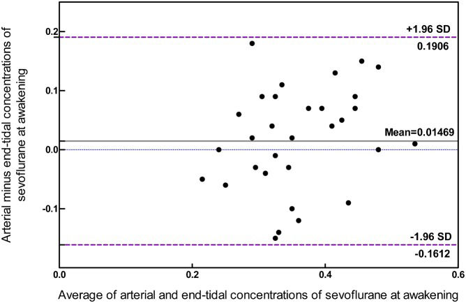
The Bland-Altman plot (Figure 2) displays the differences between the arterial concentration and end-tidal concentration at awakening. The differences between the two concentrations ranged from -0.15 to 0.18%, with a mean (SD) of 0.01% (0.09). After grouping, the differences fell into the acceptable range (±1.96 SD), regardless of the length of the awakening time.

The Bland-Altman plot displays the differences between the arterial and end-tidal concentrations at awakening plotted against the average of the two concentrations. The differences ranged from -0.15 to 0.18, all of which were within the SD range of ±1.96. The two methods may be used interchangeably.
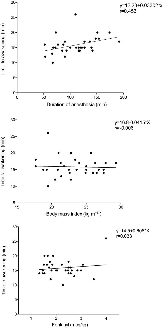
Figure 3 depicts that the time to awakening was prolonged according to the duration of anesthesia from 52 to 192 minutes (P = 0.006) but was not correlated with the body mass index (range 18-29) or the total fentanyl dose (P = 0.971 and 0.850, respectively).
DISCUSSION but was not correlated with the body mass index (P = 0.971) or total fentanyl dose (P = 0.850).")
This study first quantitatively demonstrated that the awakening arterial blood concentration of sevoflurane was 0.36% (0.10) in surgical gynecologic patients. An increased duration of sevoflurane anesthesia within 3 hours did not elevate the arterial concentration before discontinuing sevoflurane but did prolong the awakening time. With well-assisted ventilation, the end-tidal concentration of sevoflurane was nearly equal to the arterial concentration and could be a reliable predictor for eye opening during the emergence from general anesthesia.
The total body uptake and elimination of inhaled anesthetics should be proportional to the duration of general anesthesia 18,19. Compared with desflurane, the higher blood-gas and tissue-blood partition coefficients of sevoflurane theoretically increase total body uptake and detain elimination in patients with a longer anesthetic duration and higher body mass index 7,20. Mckay et al. 7 demonstrated that the time from anesthetic discontinuation to the first response to verbal command and the time to recover the ability to swallow after sevoflurane exceeded those after desflurane. In addition, the contribution of the body mass index to this delay of airway reflex recovery was more pronounced after sevoflurane anesthesia. Sevoflurane has been clearly proven to prolong the extubation time compared with desflurane (20-25%) 6 but to reduce the mean extubation time by 13% relative to isoflurane 21. In our study, the arterial concentration before discontinuing sevoflurane was not correlated with the duration of anesthesia (55-192 min), indicating that a limited content of sevoflurane was present in the blood (lower blood-gas partition coefficient, 0.65). However, the time to awakening was proportionally prolonged by a longer duration of anesthesia, further indicating increased body uptake (higher muscle-blood and fat-blood partition coefficients, 3.1 and 47.5, respectively) 5. By measuring arterial blood concentrations during the emergence period, this study provided convincing evidence for sevoflurane elimination that should be taken into consideration in cases of a prolonged duration of anesthesia.
The rate of uptake of inhaled anesthetics depends on the alveolar concentration and ventilation 22-25, blood solubility 5 and cardiac output 22. Similarly, sevoflurane washout commences from the brain and body via the circulating blood into the alveolar space during elimination, allowing the lungs to ventilate it into the air. When the brain or arterial concentration decreases to a certain level, the patient awakens enough to follow the verbal command for eye opening. In this study, the end-tidal concentrations of sevoflurane decreased more prominently in the initial 5 minutes after discontinuation, indicating a rapid alveolar washout, and then became consistently close to the arterial concentrations. Our previous study demonstrated that the end-tidal concentrations of desflurane are generally lower than the arterial blood concentrations during elimination in gynecologic patients 8; this effect is related to the lower blood-gas partition coefficient of 0.42 and tissue/blood partition coefficients of desflurane 5. By determining the arterial blood concentration, we therefore identified that the end-tidal concentration of sevoflurane, which is close to 0.36%, is a reliable indicator for predicting emergence from general anesthesia.
There are two limitations in the current study. First, all of our patients were female, had BMIs less than 29 and received a maximum 3-hour duration of anesthesia. The minimum alveolar concentration (MAC) value of sevoflurane varied from 2.6% in young adults (age 18-35 yrs) to 1.58% to 2.05% in middle-aged adults (age 16-59 yrs), to 1.45% in elderly people (age over 70 yrs) 1. The MAC-awake of sevoflurane in children ranged from 0.43 to 0.66% 26, whereas the awakening time was significantly shorter in male patients than in females 27. In our study, the relatively lean body size and shorter duration of general anesthesia may not demonstrate the BMI effect of sevoflurane compared with previous studies 7,20. The clinical inference should be adjusted for the patient's characteristics. Second, the minute ventilation could hardly be controlled by a ventilator after the reversal of spontaneous breathing during emergence. We therefore manually assisted the ventilation to keep the end-tidal CO2 concentrations as close to 38-42 mmHg as possible before extubation. The end-tidal CO2 depicts the status of minute ventilation and cardiac output. Hyperventilation may decrease the PaCO2 and cerebral blood flow and then depress cardiac output, which reverses and decelerates the elimination of inhaled anesthetics. In our study, the minute ventilation and cardiac index of all patients was kept steady before extubation, which revealed a good agreement between the awakening arterial blood and end-tidal sevoflurane concentrations. By investigating both arterial blood and end-tidal concentrations, the clinical influence of minute ventilation and cardiac output can be further clarified during the elimination of inhaled anesthetics.
In conclusion, we have demonstrated the awakening arterial concentration of sevoflurane in gynecologic patients. An increased duration of sevoflurane anesthesia (1-3 hours) did not significantly elevate arterial concentrations before discontinuation and at awakening but did prolong the time to awakening, indicating limited blood uptake but increased body uptake of sevoflurane. With well-assisted ventilation during emergence, the end-tidal concentration of sevoflurane can be a reliable indicator for predicting awakening from general anesthesia.
AUTHOR CONTRIBUTIONSLin TC, Lu CC, and Hsu HC performed this clinical study and drafted the manuscript. Lee MS performed the statistical analysis. Ho ST conceived the study and provided laboratory support to measure the blood concentrations. Su HY participated in the study design and coordination. All authors read and approved the final version of the manuscript.
We gratefully acknowledge Ms Yi-Fon Roa, who assisted with the gas chromatography headspace sampler system for blood sevoflurane determination, and the research assistant, Ms Yi-Ru Chen. This study was funded by a grant (NSC98-2314-B-016-007-MY3) from the Taiwan National Council of Science. This work was performed in the Tri-Service General Hospital/National Defense Medical Center in Taipei, Taiwan.
No potential conflict of interest was reported.