


This study investigated the acute hemodynamic responses to multiple sets of passive stretching exercises performed with and without the Valsalva maneuver.
METHODS:Fifteen healthy men aged 21 to 29 years with poor flexibility performed stretching protocols comprising 10 sets of maximal passive unilateral hip flexion, sustained for 30 seconds with equal intervals between sets. Protocols without and with the Valsalva maneuver were applied in a random counterbalanced order, separated by 48-hour intervals. Hemodynamic responses were measured by photoplethysmography pre-exercise, during the stretching sets, and post-exercise.
RESULTS:The effects of stretching sets on systolic and diastolic blood pressure were cumulative until the fourth set in protocols performed with and without the Valsalva maneuver. The heart rate and rate pressure product increased in both protocols, but no additive effect was observed due to the number of sets. Hemodynamic responses were always higher when stretching was performed with the Valsalva maneuver, causing an additional elevation in the rate pressure product.
CONCLUSIONS:Multiple sets of unilateral hip flexion stretching significantly increased blood pressure, heart rate, and rate pressure product values. A cumulative effect of the number of sets occurred only for systolic and diastolic blood pressure, at least in the initial sets of the stretching protocols. The performance of the Valsalva maneuver intensified all hemodynamic responses, which resulted in significant increases in cardiac work during stretching exercises.
Flexibility is considered a component of physical fitness that is related to health, and flexibility training is recommended in different contexts 1. Recommendations regarding flexibility training include at least four sets of exercises for large muscle groups, performed two or three times per week 2. To achieve positive results and avoid overload, movements should not exceed the point of discomfort 1,2.
Muscle contraction during stretching exercises might increase hemodynamic responses 3,4, which appear to be influenced by the amount of activated muscle mass 5,6 and the intensity of stretching 6. These changes are primarily mediated by type III afferent fibers, which are sensitive to mechanical stress. The stimulation of these peripheral mechanoreceptors induces vagal withdrawal and increases sympathetic activity 7,8,9.
Although previous studies have shown that acute stretching exercise induces transient cardiovascular responses, the pattern of these responses remains controversial. Some studies have observed an increase in arterial blood pressure (BP) 10, heart rate (HR) 3,11, or both 12,13,7,8,4. However, most of these studies primarily focused on mechanisms associated with these variations, neglecting aspects related to exercise prescription variables. From a practical perspective, studying these variables is important because variables such as the set number, length stimulus, interval between sets, and training method (dynamic or static) likely affect the hemodynamic responses to acute stretching exercise 10,5,6.
The influence of the Valsalva maneuver (VM) during flexibility training routines also requires investigation because cardiovascular responses are probably greater when it is performed 14. This maneuver might frequently be used during stretching exercises, especially in cases of less flexible individuals, due to difficulty reaching and sustaining extreme ranges of motion for several seconds 5. A previous study conducted by our group assessed the influence of four sets of stretching exercises performed with and without the VM on acute cardiovascular responses 5. Both the BP and rate pressure product increased up to the fourth set, especially when the exercises were performed with larger (hamstrings) vs. smaller (gastrocnemius) muscle groups and with the VM. However, in this prior study, the BP was measured manually, which might have affected data precision. Furthermore, current recommendations for health-related exercise indicate that a higher number of sets should be performed during flexibility training 2. Therefore, the acute hemodynamic responses induced by multiple successive sets of stretching exercises should be investigated. This information might be particularly useful for stretching routines designed for populations at cardiovascular risk.
Considering the lack of studies investigating acute cardiovascular responses to multiple sets of stretching exercises performed with and without the VM and the importance of these aspects for exercise prescription, the objective of this study was to compare the acute responses of blood pressure, HR, and rate pressure product during 10 sets of passive static stretching of the hip joint performed with and without the VM. It has been hypothesized that the number of sets would have a cumulative effect upon the hemodynamic responses and that performance of the VM would induce further increases in all observed variables.
MATERIALS AND METHODSExperimental approach to the problemData were collected during three laboratory visits. On the first day, the informed consent form was signed, the Physical Activity Readiness Questionnaire (PAR-Q) was administered, and anthropometric and flexibility measures were conducted. A flexibility assessment was necessary to assure sample homogeneity with regard to maximum joint range of motion (ROM). Maximal passive flexibility was measured by means of a portable angular device (Fleximeter™, Campinas, SP, Brazil). An increase in hemodynamic responses would be more likely to occur in individuals with poor flexibility due to greater static contraction performed at the extreme ROMs typically applied during this mode of flexibility training 15,16; thus, cut-off points were applied to include only subjects with low flexibility levels in the study.
On the second and third visits, which were separated by an interval of at least 48 hours, systolic and diastolic BP (SBP/DBP) and HR were continuously measured by infrared photoplethysmography (Finometer Pro™, Finapres Medical Systems, Amsterdam, The Netherlands) for 10 minutes at rest, during 10 sets of stretching exercise, and for 10 minutes post-exercise. The rate pressure product was calculated as the product of the HR and SBP. The multiple sets of unilateral hip flexion stretching exercise were performed with or without the VM on different days, defined in a counter-balanced order. Individuals were instructed to avoid practicing any type of physical activity 24 hours prior to the experimental sessions.
SubjectsThe sample comprised 15 asymptomatic men (mean±standard deviation; age 23.6±2.8 years; body mass index 24.2±4.0 kg/m2; resting HR 68.0±8.8 bpm; SBP 126.0±9.2 mmHg; DBP 70.0±4.6 mmHg; and hip flexion ROM 84.9±9.4°) with no previous flexibility training experience. Eligible subjects exhibited a low flexibility level, defined as a maximum ROM of 90° for the unilateral hip flexion exercise 16. Additionally, the following exclusion criteria were adopted: use of medications that could potentially influence the hemodynamic responses to exercise; bone, joint, or muscle problems limiting unilateral hip flexion performance; and positive PAR-Q results.
Stretching exercisesWhile performing the stretching exercise, the volunteers were instructed to reach their maximum ROM, and acute hemodynamic variables were continuously measured. The passive static stretching method was applied with four sets of 30-second stimuli and 30-second intervals between sets. For exercises performed with the VM, breathing was voluntarily interrupted during the last 15 seconds of the stretching exercise.
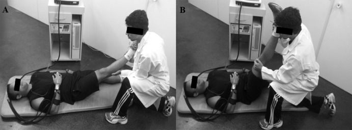
Unilateral hip flexion exercises were standardized as follows: a) For the initial and final position, the subject remained in a supine position, lying on a mat. b) For the intermediate position, the subject remained with slight hip flexion of the dominant leg for 25 seconds (Figure 1A). The intent of this procedure was to reduce the time between the beginning of the motion and reaching the maximal ROM. c) For the maximum intensity position, unilateral hip flexion was conducted by moving the dominant leg with the knee extended to the maximum ROM while the other limb remained extended and in contact with the ground. The maximum ROM was determined as the point associated with the subject's subjective indication of pain sensation or mechanical restriction to the movement. The ankle was free to prevent tension from being transmitted through the triceps surae, and the knees were extended during the entire movement (Figure 1B).
 before moving the segment to the maximum range of motion; and (B) at the maximum range of motion. Evaluators responsible for stabilizing the hip joint and measuring the duration of stretching and intervals between sets are not shown for the sake of clarity.")
Unilateral hip flexion stretching exercise: (A) before moving the segment to the maximum range of motion; and (B) at the maximum range of motion. Evaluators responsible for stabilizing the hip joint and measuring the duration of stretching and intervals between sets are not shown for the sake of clarity.
Although only one evaluator manipulated the segment, three evaluators were required to execute the entire stretching protocol. The first evaluator executed the stretching protocol. The second evaluator was responsible for controlling the stretching duration and the intervals between sets. Finally, the third evaluator held the contralateral hip to stabilize the movement.
Hemodynamic assessmentThe hemodynamic variables were measured at three different times, always in the dominant limb, as follows: at rest, during the stretching exercise, and during post-exercise recovery. All measurements were performed with the subjects laying in a supine position on a mat in a silent, thermoneutral environment (mean±SD: 22±1°C). HR, SBP and DBP were assessed by finger photoplethysmography (FinometerTM, Finapres Medical Systems, Amsterdam, The Netherlands). Resting data were monitored for 10 minutes; values from the first five minutes were used for device calibration, and values from the last five minutes were averaged for the final result. Post-exercise assessment data included values averaged for five and ten minutes of recovery. Hemodynamic variables were averaged every five seconds during the stretching exercise, and the highest mean value obtained within the last 15 seconds of each set was adopted as the outcome for that set.
Statistical analysesData normality was confirmed by the Kolmogorov-Smirnov test, and the data were expressed as the mean±standard deviation. Differences between hemodynamic variables assessed at rest, during the stretching protocol, and during post-exercise recovery were tested by 2-way repeated measures analysis of variance (ANOVA). Fisher post hoc tests were used to locate differences in the case of significant F ratios (p≤0.05). All calculations were made using StatisticaTM 7.0 software (Statsoft, Tulsa, OK, USA).
EthicsPrior to participating in the study, volunteers signed a free and informed consent form, and this study was conducted according to the institutional guidelines and Resolution 96/196 from the National Council of Health/Brazil, based on the Declaration of Helsinki. The study was approved by the Rio de Janeiro State University institutional ethics committee (ethics approval 113/2011).
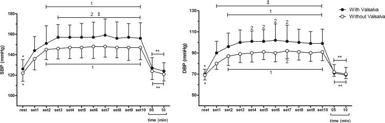
RESULTSThe results for BP at rest, during the stretching protocols, and post-exercise are displayed in Figure 2. Both SBP and DBP were higher during all stretching sets compared with the values obtained at rest and post-exercise (p≤0.001). Overall, BP increases were cumulative from the first set to the third/fourth sets (p≤0.04) and stabilized during the additional sets (p>0.05). Finally, SBP and DBP responses were systematically higher (by approximately 10-12 mmHg) when the stretching protocol was performed with the VM vs. without the VM (p<0.03 and p<0.04, respectively).
 and diastolic blood pressure (DBP) at rest, during stretching protocols performed with and without the Valsalva maneuver, and at 10 min post-exercise. Superscripted numbers represent significant differences vs. the indicated set. *: significant difference between the resting condition and all sets (p<0.05). **: Significant difference between post-exercise assessments and all sets (p<0.05). ‡: Significant difference between sets in protocols performed with the VM vs. without the VM (p<0.05).")
Systolic blood pressure (SBP) and diastolic blood pressure (DBP) at rest, during stretching protocols performed with and without the Valsalva maneuver, and at 10 min post-exercise. Superscripted numbers represent significant differences vs. the indicated set. *: significant difference between the resting condition and all sets (p<0.05). **: Significant difference between post-exercise assessments and all sets (p<0.05). ‡: Significant difference between sets in protocols performed with the VM vs. without the VM (p<0.05).
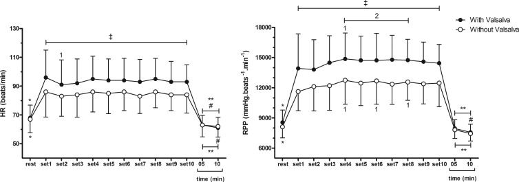
Figure 3 depicts HR and rate pressure product data. In contrast to BP, HR responses did not appear to be cumulatively affected by successive stretching sets. In fact, after an initial increase in the first set vs. pre-exercise (p≤0.01), HR values remained stable during the remaining 10 sets. However, similarly to what happened with the BP responses, stretching performed with the VM elicited greater HR increases during all sets compared with the protocol performed without the VM (p≤0.001). Rate pressure product responses appeared to be more closely related to HR than SBP. In fact, no additive effect of consecutive stretching sets could be detected for this marker of cardiac load (p>0.05), but the rate pressure product values were approximately 20% higher with the performance of the VM during all sets (p≤0.02).
 and rate pressure product (RPP) at rest, during stretching protocols performed with and without the Valsalva maneuver, and at 10 min post-exercise. Superscripted numbers represent significant differences vs. the indicated set. *: Significant difference between the resting condition and all sets (p<0.05). **: Significant difference between post-exercise assessments and all sets (p<0.05). ‡: Significant difference between sets in protocols performed with the VM vs. without the VM (p<0.05).")
Heart rate (HR) and rate pressure product (RPP) at rest, during stretching protocols performed with and without the Valsalva maneuver, and at 10 min post-exercise. Superscripted numbers represent significant differences vs. the indicated set. *: Significant difference between the resting condition and all sets (p<0.05). **: Significant difference between post-exercise assessments and all sets (p<0.05). ‡: Significant difference between sets in protocols performed with the VM vs. without the VM (p<0.05).
The present study investigated the hemodynamic responses within 10 sets of passive static stretching of the hip joint performed with and without the VM. The main findings were as follows: a) A cumulative effect of successive stretching sets was observed for BP, at least until the third/fourth sets. Both SBP and DBP appeared to stabilize during the additional sets. b) HR and rate pressure product values increased during the stretching protocols, but no additive effect related to multiple successive sets could be detected. c) The hemodynamic responses were systematically heightened by the performance of the VM, leading to an approximately 20% increase in the rate pressure product.
Our findings concur with previous studies investigating the effects of acute stretching exercise on SBP and DBP 10,5,4,6, HR 4,6, and rate pressure product 5,4. However, the present study is the first to demonstrate that a) the increase in BP appears to be cumulatively affected by the number of sets of a stretching exercise that are performed, but this additive effect appears to stabilize after the fourth set; and b) this additive effect does not appear to influence the increases in HR and rate pressure product. Moreover, VM performance during acute stretching exercise potentiates the hemodynamic responses, regardless of the number of sets. These results have important practical implications, especially for prescribing exercise for populations with cardiovascular constraints.
Some mechanisms have been suggested to mediate the increase in hemodynamic responses during stretching exercises. Gladwell and Coote 3 reported a transitory increase in HR at the start of stretching, concomitant to a decrease in parasympathetic activity 3, whereas Farinatti et al. 15 suggested that the increase in HR during stretching would be related to higher sympathetic activation and vagal withdrawal 15. In the present study, the increase in HR within the first seconds of stretching probably occurred due to a combination of mechanoreceptor stimulation and decreased baroreflex sensitivity 17. However, after the initial stimulation, an accommodation of stretched collagen molecules and a reduction in muscle spindle drive are expected, which probably reduced the ergoreflex compared with the beginning of the exercise 18. This mechanism, combined with a relatively short period of stimulation (30 sec), would help explain the absence of a cumulative effect of multiple stretching sets on the HR.
Additionally, increases in hemodynamic responses during stretching are probably mediated by neural impulses generated by mechanical activity and transmitted by muscle receptors, particularly type III afferent fibers 9,19, which are activated by both stretching and static contraction 20. The afferent influx generated by these fibers is capable of inducing increases in cardiovascular responses, even during passive stretching 21. During active stretching performed by individuals with poor flexibility, higher muscle tension is probably needed to reach and sustain a given ROM 15,22. Therefore, a greater stimulation of type III afferent fibers might occur in subjects with lower vs. higher flexibility levels. This potential mechanism could at least partially explain the increase in hemodynamic responses presently observed.
Mechano- and metaboreceptor activation during stretching exercises performed at maximum ROM might also induce baroreflex stimulation and vagal inhibition, contributing to increased hemodynamic responses 12,23. In a previous study 13, 12 healthy subjects performed 25 repetitions of 5 seconds of calf muscle stretching interspersed with periods of 15 to 25 seconds of recovery. Using this protocol, isolated mechanoreceptor stimulation was capable of increasing variables such as HR, sympathetic activity, and SBP, albeit temporarily and with a small latency period at the beginning of stretching. While stimulation via baroreceptors was rapidly normalized, the increase in hemodynamic responses would likely be sustained longer by an afferent influx from metaboreceptors.
The present data indicated that VM performance during stretching potentiated the increase in all hemodynamic responses. The VM is classically defined as compulsory exhalation against a closed glottis, thereby increasing the thoracic pressure and precluding venous return of blood to the heart 24. Typical arterial pressure and HR changes include a brief increase in BP due to the propulsion of blood from the thorax, followed by a baroreflex-mediated reduction and posterior elevation produced by a combination of vasoconstriction, sympathetic cardiac stimulation, and tachycardia 25. Hence, occurrence of the VM during any type of exercise would be expected to increase the hemodynamic responses, which has been confirmed in this study.
The novelty of our findings pertains to the extent to which VM performance during stretching exercise specifically impacted BP and cardiac work in individuals with poor flexibility levels. The rate pressure product is known to reflect cardiac work during strength and aerobic exercises 26. However, little information exists in the literature regarding its responses to stretching exercises. We have demonstrated that VM performance increased the rate pressure product response by 20% in every one of 10 sets of passive unilateral hip flexion stretching 27,28. Such a response calls attention to the fact that the cardiovascular stress to overcome mechanical and antagonist resistance during stretching exercises is not negligible. This must be considered to improve safety during training sessions, which usually include several exercises involving large muscle groups and active static contraction to hold the movement at extreme ROMs. Additional studies are therefore warranted to confirm the present results and to better describe hemodynamic responses during flexibility training sessions comprising several exercises, as is the case in actual prescribed sets.
The major limitation of this study concerned the lack of control of muscular activity and of the pressure or lung volume during the VM. Although volunteers were instructed to relax the quadriceps during hip flexion, this was not confirmed by means of electromyography monitoring, which would help to detect involuntary muscle contraction. The absence of data about the actual pressure or lung volume during the VM precluded the observation of the isolated effect of the VM on BP. Notably, subjects might have performed the maneuver with different volumes and pressures, which could have influenced the hemodynamic responses. However, in real training sets, these variables are not controlled, and we tried to reproduce what is actually observed during stretching routines. Because subjects were quite homogeneous with regard to age and physical fitness level, possible variations in VM performance likely did not compromise our results. Finally, the extent to which our data can be applied to training sessions that include multiple sets of various exercises must be defined by further research.
In conclusion, multiple sets of the passive stretching of unilateral hip flexion exercise performed for 30 seconds significantly increased the BP, HR, and rate pressure product. The number of sets had a cumulative effect on SBP and DBP but not on HR and rate pressure product. VM performance for the last 15 seconds of stretching sets consistently intensified all hemodynamic responses, resulting in an additional increase of approximately 20% in the cardiac work, as estimated by the rate pressure product.
AUTHOR CONTRIBUTIONSLima TP and Moteiro WD designed the study and contributed to the data analysis and interpretation and manuscript drafting and review. Farinatti PT, Rubini EC and Silva EB contributed to the data analysis and interpretation and manuscript drafting and review. All of the authors declare that they participated sufficiently in the work to take public responsibility.
This study was partially supported by grants from the Brazilian Council for Research and Technological Development (CNPq) and the Carlos Chagas Foundation for Research Support in Rio de Janeiro (FAPERJ).
No potential conflict of interest was reported.