


Metanephric adenoma (MA), also designated nephronogenic nephroma or renal epithelial tumor resembling immature nephron, has just been recently recognized as a special type of benign renal epithelial tumor. Only few reports are found in the literature regarding this rare renal tumor. The purpose of this paper is to describe our clinical, imaging and histological / immunohistochemical observations of MA diagnosed in two patients and compare these data to previous information reported in medical databases.
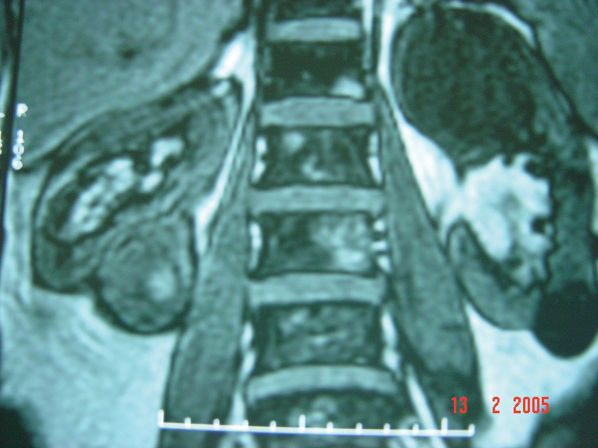
Case ReportThe first case refers to a 70-years-old female patient, who except by systemic arterial hypertension was completely healthy. During routine examinations, a mass of 4.5 cm located on the lower third of her right kidney was incidentally found. At that time, the patient presented only low-grade fever and weakness for about two weeks. Abdominal ultrassonography (USG) showed hypoechoec mass with solid aspect and irregular limits. It was heterogeneous and exofitic, containing septation and cystic areas in its interior. Magnetic resonance imaging (MRI) showed a solid mass 5.2 cm for its higher dimension located in right kidney with same hemorrhagic foci and no sign of peri-renal invasion (Figure 1). Also, simple renal cysts were evidenced. The patient was submitted to a successful partial nephrectomy by open surgery, evolving uneventfully. Pathologic examination of surgical specimen revealed a firm lesion of light brown tissue with reticulated central area and clear delimitation from the adjacent parenchyma. Microscopic evaluation showed no cellular atypical or mitotic activity. Peri-renal adipose tissue was free from neoplasic invasion.

The second patient was a 23-year-old man with no previous heath problem, who presented with flank pain for one month. Abdominal USG showed a 4×4 cm nodular, solid and heterogenic lesion with hyperecogenic areas and exofitic characteristics in the lower third of right renal parenchyma. Computed tomography (CT) confirmed the presence of a 4 cm expansive tumoral formation with intermediate attenuation and minimum venous contrast enhancing at that anatomic site (Figure 2). Partial nephrectomy was again the therapeutic choice. The patient evolved well but an arterial-venous fistula clinically manifested by hematuria was diagnosed in post-operative (PO) day five. The fistula was successfully embolized by arteriography and no other complications occurred. Immunohistochemical study employing incubation of histological cuts with mono and polyclonal antibodies (panels) showed a profile focally and positive to WT-1, EMA and CK7.

Both patients are free of neoplasic disease with no signs of recurrence after 2 years of follow up.
DISCUSSIONMA is a renal tumor with benign behavior. The classification is based on a combination of histological, immunohistochemical, and genetic features. Most small MA tumors are well circumscribed, firm, and white; larger tumors may be hemorrhagic and softer.
The cytology of a few cases has been described1 and is composed of tight, short papillae and loose sheets of cells with scant cytoplasm, round nuclei, fine chromatin and rare small nucleoli. By analyzing all described findings at immunohistochemical and lectin histochemical studies, MA has shown reactivity for keratin (CK7 is the most common), CD 57, vimentin, S-100 protein, EMA, lysozyme, a-1-antitrypsin, PNA, DBA, SBA and WT-1.2 It seems that, despite the variability, CK7 focally positive is a rule. In our second case, immunohistochemic examination revealed WT-1, EMA and CK7 locally positive.
Unlike renal adenoma, which is by definition <5 mm in diameter, MA can grow to a large size. The diameter of MA ranges from 6 to 200 mm2. Our findings were renal tumors of 40 and 45 mm. All MA reported, except one,3 behaved in a benign way with no metastatic findings or local recurrence after surgical removal, showing no evidence of cellular atypical or mitotic activity.2 Similar fact was observed for our two patients. For their special findings in MA variants, some authors deserve brief comment. In 2000, Renshaw et al 3 presented the only reported case of a metastatic disease from a typical MA occurring in a 7 year old child, but no death related to the tumor has been demonstrated.
Clinical manifestations related to MA are very unspecific and sometimes even absent, being the majority of these renal tumors incidentally found.4 In 1995, four of eight cases reported by Nonomura et al5 were found with no signs or symptoms and two presented only a tumoral mass. In the same year, Jones et al6 reported one tumor found only at autopsy and six tumors discovered during other pathological investigation. When present, signs and symptoms include abdominal or flank pain, hematuria, palpable mass, hypertension and fever. Our cases presented some of these clinical findings.
At present there are only few reports regarding the imaging findings of MA. Some authors found adenomas to be hyperechoic at USG.7,8 MA has been described to show enhancement on contrast CT, but angiography reveals neither neovascularization nor tumor staining. 7,8 On both T1- and T2-weighted MR images, the tumor is represented as an isointense mass.5,7 On the imaging findings of MA, hypovascularity and frequent calcification may be rather characteristic. Bastide et al9 reported their imaging findings in nine patients, describing MA as a lesion with no vascular flow on color Doppler USG, presence of calcifications, and minimal enhancement in contrast CT. It is possible to realize that MA has same common findings, however none is so specific neither can exclude malignity.
Most reports describe total nephrectomy as gold standard treatment for MA, but partial nephrectomy must be considered a good option. Nowadays, it is known that partial nephrectomy is indicated even to malignant tumors and even large masses (bigger than 4 cm) can be ressected without prejudice to the oncological results. In 2000, Kosugi et al.10 performed partial nephrectomy for a 32 years old female patient who presented with a right renal MA of 1.5 cm at abdominal USG examination at a routine medical check up. Our two patients were also treated with partial nephrectomy, procedure with better renal function preservation, without compromising the patient's survival. Further advance in reducing patient morbidity may be achieved by laparoscopic nephrectomy as reported by Ebine et al.11 in 2004 for MA treatment in a 31 years old female patient with a left renal mass of 4.5 cm detected incidentally during an abdominal ultrasound examination. Three years latter, Kumar et al12 reported a laparoscopic partial nephrectomy as treatment for MA in a 47-year-old patient. In the biggest MA series reported, Bastide et al9 performed four radical and five partial nephrectomies. In this paper, authors suggest a preoperative diagnostic biopsy, a partial nephrectomy or active surveillance. We believe, that biopsy should be reserved to cases where patient refuse or does not have clinical conditions for surgery or less invasive procedure shall be tried, such as cryotherapy.
It is possible to realize that MA treatment has evolved as well as others renal tumors approach, and partial nephrectomy should be remembered as an option (Table 1).