metricas
covid
Buscar en
Gastroenterología y Hepatología (English Edition)
Toda la web
Inicio Gastroenterología y Hepatología (English Edition) Oesophageal intramural pseudodiverticulosis, a condition to be aware of
Journal Information
Vol. 45. Issue 5.
Pages 373-374 (May 2022)
Vol. 45. Issue 5.
Pages 373-374 (May 2022)
Image of the month
Full text access
Oesophageal intramural pseudodiverticulosis, a condition to be aware of
Pseudodiverticulosis intramural esofágica, una entidad a conocer
Visits
28
Paul Gisasola Dorronsoro
Corresponding author
, Ainara Iriarte Rodríguez, Patricia Aranzabal Aguilar
Servicio de Digestivo, Hospital de Zumárraga, Zumárraga, Guipúzcoa, Spain
This item has received
Article information
Full Text
Bibliography
Download PDF
Statistics
Figures (2)
Full Text

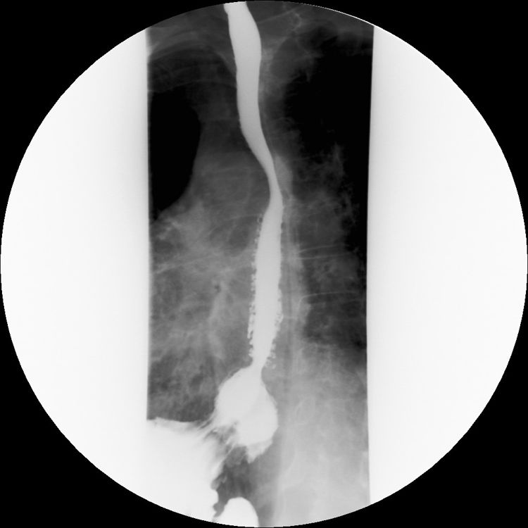
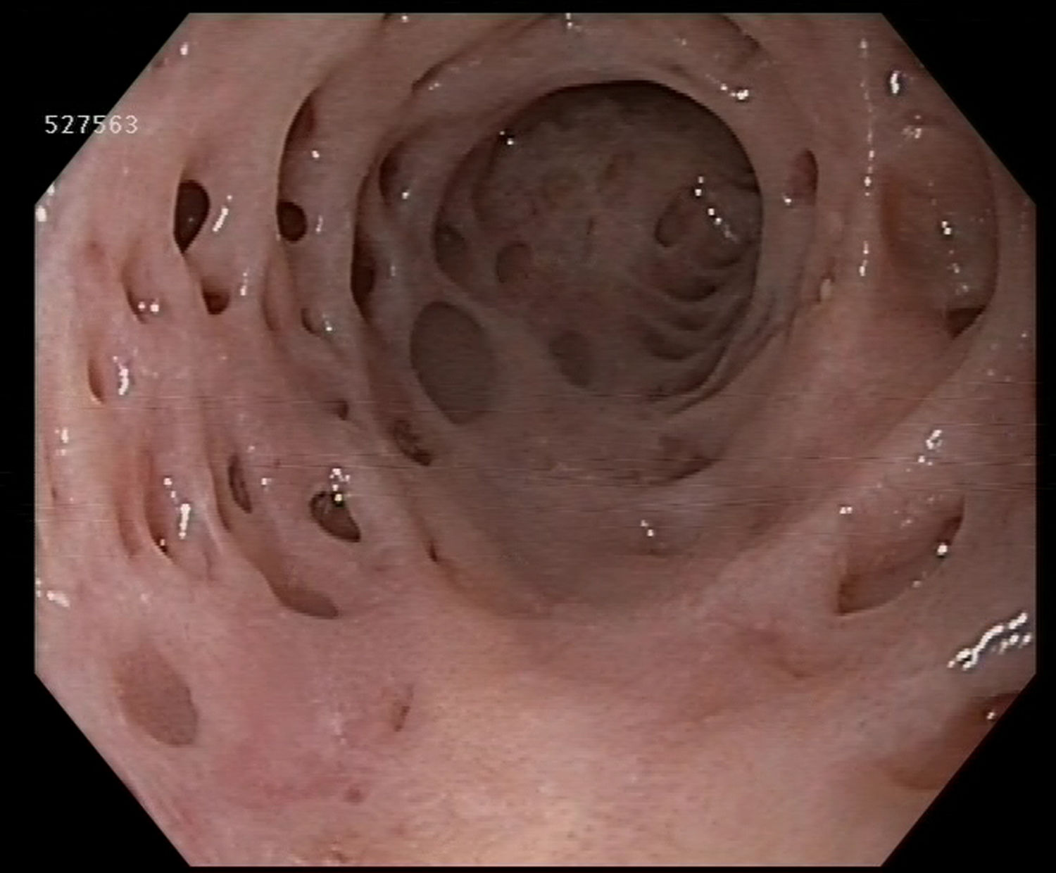
We present the endoscopic (Fig. 1) and radiological (Fig. 2) images of an oesophageal intramural pseudodiverticulosis (OIPD). It is a rare condition that occurs in fewer than 1% of upper gastrointestinal endoscopies (UGE) and it occurs as a result of hypertrophy of the submucosal mucous glands with cystic dilation of their excretion ducts. As such, it is not a true diverticulosis. With an unknown aetiology, it is associated with gastroesophageal reflux, oesophageal candidiasis and chronic alcohol use; conditions that our patient did not present. Although dysphagia is reported as the most common symptom, our 65-year-old male patient did not present with any symptoms, and the reason for the UGE was anaemia testing.2,3

Figure 1.

Oesophagogram: multiple images of pseudodiverticula.

(0.03MB).
Figure 2.

Endoscopic image of the oesophageal pseudodiverticulosis.

(0.13MB).

Small diverticula (<5 mm) may occur as either diffuse or localised, as was the case in our patient whose lesions were all in the distal segment of the oesophagus. Reported accompanying lesions include peptic stenosis and oesophageal candidiasis, which were not observed in our patient. However, he did present with an oesophageal papilloma, removed with forceps and with a confirmatory biopsy.2,3

The treatment depends on the patient's symptoms. In our case, it was decided to not start any treatment as he had no symptoms and there were no associated lesions.1,2

References
[1]
R. Plaza, A. Barreiro, R. Lorente, G. Carrión, Á. Ponferrada, M. Aldeguer.
Pseudodiverticulosis esofágica intramural: una causa poco frecuente de disfagia.
Rev Esp Enferm Dig, 106 (2014), pp. 411-412
[2]
Y.E. Chon, S. Hwang, K.S. Jung, H.J. Lee, S.G. Lee, S.K. Shin, et al.
A case of esophageal intramural pseudodiverticulosis.
Gut Liver., 5 (2011), pp. 93-95
[3]
S. Sabanathan, F.D. Salama, W.E. Morgan.
Oesophageal intramural pseudodiverticulosis.
Thorax., 40 (1985), pp. 849-857

Please cite this article as: Gisasola Dorronsoro P, Iriarte Rodríguez A, Aranzabal Aguilar P, Pseudodiverticulosis intramural esofágica, una entidad a conocer, Gastroenterología y Hepatología. 2022;45:373–374.

Copyright © 2020. Elsevier España, S.L.U.. All rights reserved
Article options
es en pt

¿Es usted profesional sanitario apto para prescribir o dispensar medicamentos?

Are you a health professional able to prescribe or dispense drugs?

Você é um profissional de saúde habilitado a prescrever ou dispensar medicamentos