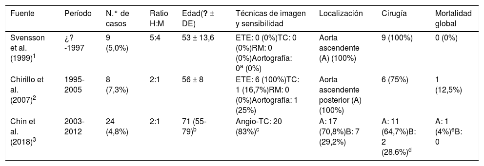
La disección aórtica es la patología aguda más frecuente de la aorta y presenta una elevada mortalidad, por lo que constituye una urgencia radiológica de vital importancia. Actualmente se diferencian cinco subtipos, siendo la clase 3, también conocida como disección aórtica limitada o sutil, la variante más desconocida. Este tipo de disección es infrecuente y exige un conocimiento claro de su semiología radiológica para que no pase desapercibida. Desde el punto de vista de la imagen, esta entidad se caracteriza por un pequeño abultamiento focal del contorno aórtico y/o dilatación circunferencial localizada del segmento aórtico afectado por el desgarro intimal. Recientemente se ha destacado la baja familiaridad del radiólogo con esta patología. Con la finalidad de ilustrar los principales hallazgos de imagen y revisar los aspectos más relevantes de esta entidad, presentamos cuatro casos de disección aórtica de clase 3 diagnosticados en nuestro hospital.
Aortic dissection (AD) is the most common acute condition of the aorta and has a high mortality. Therefore, it is a radiological emergency of vital importance. Currently, five subtypes are distinguished, among which AD class 3 –also known as limited or subtle AD- is the less recognised. This type of dissection is infrequent and needs to be acknowledged radiologically in order not to go unnoticed. Regarding its imaging features, this entity is characterized by a small focal bulging of the aortic wall outline and/or a limited round dilation at the region affected by the intimal tear. Recently, the low familiarity of the radiologist with this condition has been emphasized. With the aim of illustrating the main imaging findings of this entity and reviewing its most relevant aspects, we present four cases of AD class 3 diagnosed in our hospital.


