

Suplemento “Avances en Radiología Tóracica”
More infoNuestros objetivos son describir la semiología radiológica, los datos distintivos clínico-analíticos y el pronóstico relacionados con el signo de la diana (SD) en la COVID-19. Determinar si la tomosíntesis digital torácica (TDT) mejora la capacidad diagnóstica de la radiografía.
Material y métodosEstudio retrospectivo, descriptivo de una serie de casos, unicéntrico, aceptado por nuestro comité ético. Se analizaron las características radiológicas, clínicas, analíticas y evolutivas de los pacientes con COVID-19 y SD en radiografía y TDT entre noviembre de 2020 y enero de 2021.
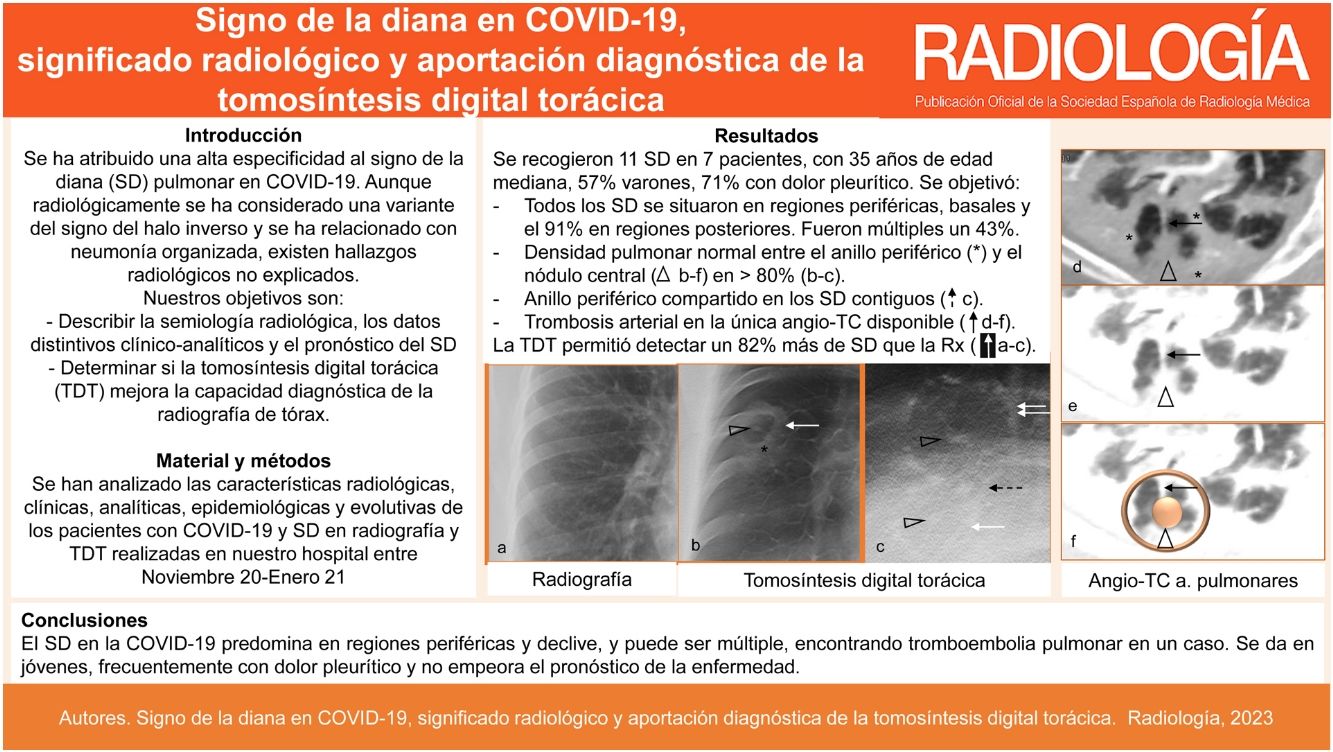
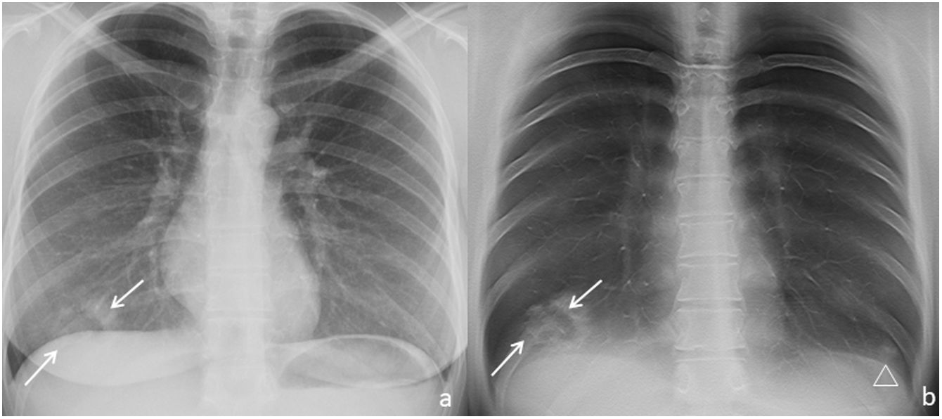
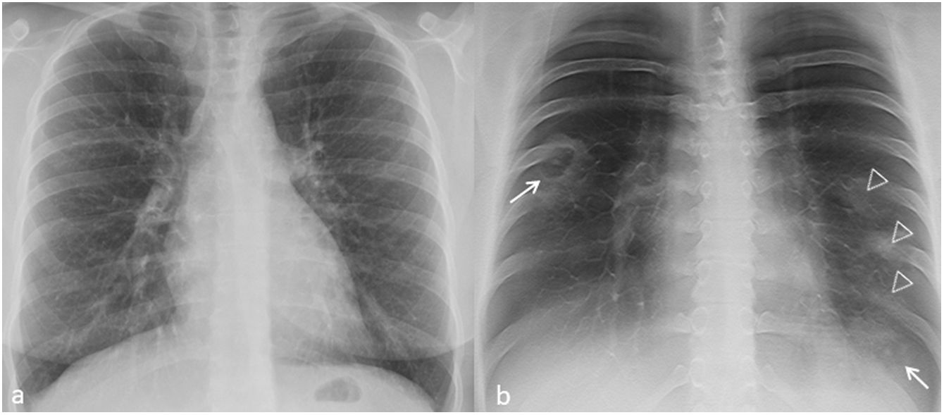
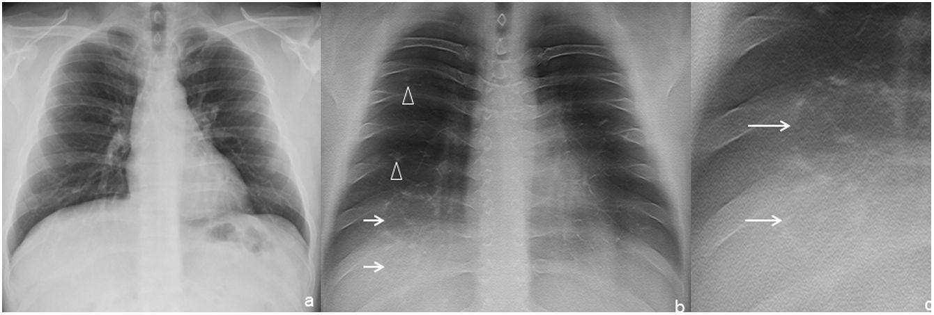
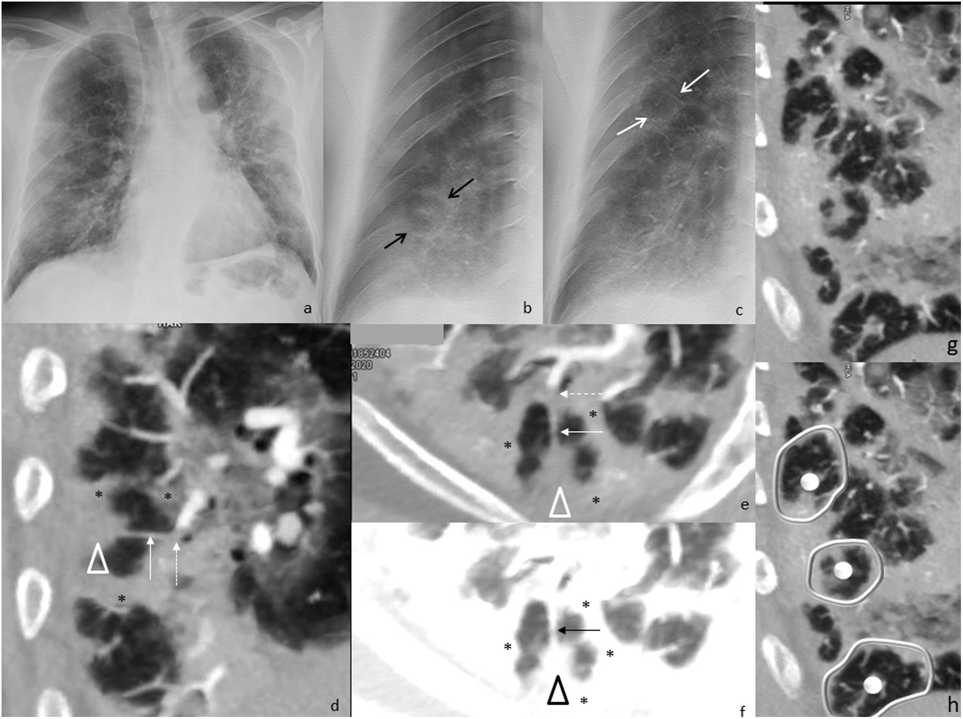
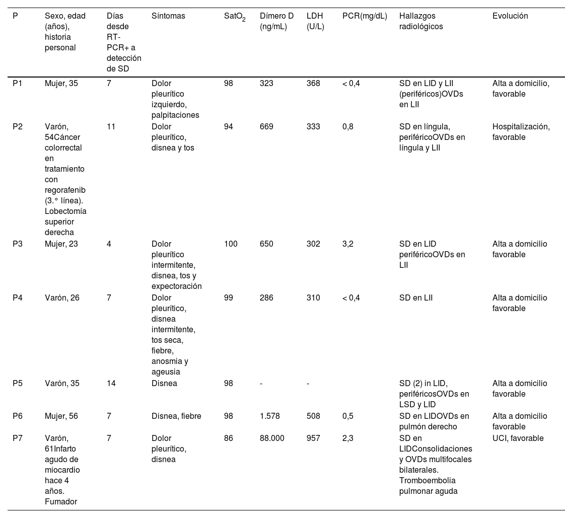
ResultadosSe recogieron 11 SD en siete pacientes, con edad mediana de 35 años, 57% varones. Todos presentaron un nódulo central y un anillo periférico y, en al menos 82%, el pulmón entre ambos fue de densidad normal. Todos se situaron en regiones periféricas, basales y 91% en regiones posteriores. Fueron múltiples en 43%. Los SD contiguos compartieron el anillo periférico; 86% asoció otras manifestaciones pulmonares de neumonía. La TDT detectó 82% más de SD que la radiografía. Solo en un paciente se realizó una angio-TC de arterias pulmonares, positiva para tromboembolia pulmonar aguda; 71% acudió con dolor pleurítico. No se detectaron hallazgos analíticos distintivos ni empeoramiento pronóstico.
ConclusionesEl SD en la COVID-19 predomina en regiones periféricas y declive y puede ser múltiple, encontrando tromboembolia pulmonar en un caso. Se da en jóvenes, frecuentemente con dolor pleurítico y no empeora el pronóstico. La TDT detecta más de 80% de SD que la radiografía.
Our objectives are to describe the imaging features, clinical characteristics, laboratory values and prognosis related to the target sign (TS) in COVID-19, as well as to determine whether digital tomosynthesis (DTS) of the chest has diagnostic advantages over radiography in this context.
Material and methodsRetrospective, descriptive, single-centre, case series study, accepted by our ethical committee. Radiological, clinical, analytical and follow-up characteristics of patients with COVID-19 and TS on radiography and DTS between November 2020 and January 2021 were analysed.
ResultsEleven TS were collected in seven patients, with a median age of 35 years, 57% male. All TS presented with a central nodule and a peripheral ring, and in at least 82%, the lung in between was of normal density. All TS were located in peripheral, basal regions and 91% in posterior regions. TS were multiple in 43%. The peripheral rings in contiguous TS were joined. Other pneumonia-related findings were identified in 86% of patients. Detection of the TS 82% higher in DTS than radiography. Only one patient underwent a CT pulmonary angiography, which was positive for acute pulmonary embolism. Seventy-one per cent presented with pleuritic pain. Laboratory values revealed no significant findings and the TS did not indicate a worse prognosis.
ConclusionsTS in COVID-19 predominates in peripheral and basal regions and can be multiple. Pulmonary embolism was detected in one case. It occurs in young people, frequently with pleuritic pain and does not lead to a worse prognosis. DTS detects TS in over 80% more cases than radiography.