Suplement “"Advances in Thoracic Radiology”
More infoMesothelioma is an infrequent neoplasm with a poor prognosis that is related to exposure to asbestos and whose peak incidence in Europe is estimated from 2020. Its diagnosis is complex; imaging techniques and the performance of invasive pleural techniques being essential for pathological confirmation. The different diagnostic yields of these invasive techniques are collected in the medical literature. The present work consisted of reviewing how the definitive diagnosis of mesothelioma cases in our centre was reached to check if there was concordance with the data in the bibliography.
Materials and methodsRetrospective review of patients with a diagnosis of pleural mesothelioma in the period 2019–2021, analysing demographic data and exposure to asbestos, the semiology of the radiological findings and the invasive techniques performed to reach the diagnosis.
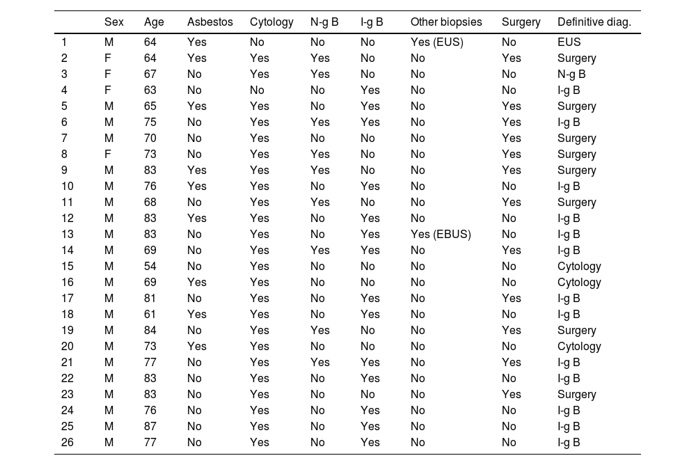
ResultsTwenty-six mesothelioma cases were reviewed. 22 men and 4 women. Median age 74 years. 9 patients had a history of asbestos exposure. Moderate-severe pleural effusion was the most frequent radiological finding (23/26). The sensitivity of the invasive techniques was as follows: Cytology 13%, biopsy without image guidance 11%, image-guided biopsy 93%, surgical biopsy 67%.
ConclusionsIn our review, pleural biopsy performed with image guidance was the test that had the highest diagnostic yield, so it should be considered as the initial invasive test for the study of mesothelioma.
El mesotelioma es una neoplasia poco frecuente y con mal pronóstico que se relaciona con la exposición al asbesto y cuyo pico de incidencia en Europa se estima a partir del 2020. Su diagnóstico es complejo, siendo fundamentales las técnicas de imagen y la realización de técnicas invasivas pleurales para la confirmación anatomopatológica. En la bibliografía médica se recogen las distintas rentabilidades diagnósticas de estas técnicas invasivas. El presente trabajo consistió en revisar cómo se llegó al diagnóstico definitivo de casos de mesotelioma de nuestro centro para comprobar si existía concordancia con los datos de la bibliografía.
Materiales y métodosRevisión retrospectiva de pacientes con diagnóstico de mesotelioma pleural en el periodo 2019–2021 analizando los datos demográficos y de exposición a asbesto, la semiología de los hallazgos radiológicos y las técnicas invasivas realizadas para llegar al diagnóstico.
ResultadosSe revisaron 26 casos de mesotelioma. 22 hombres y 4 mujeres. Edad media de 74 años. 9 pacientes tenían antecedentes de exposición al asbesto. El derrame pleural moderado-importante fue el hallazgo radiológico más frecuente (23/26). La sensibilidad de las técnicas invasivas fue de un 13% para la citología, de un 11% para la biopsia sin guía de imagen, de un 93% para la biopsia guiada por técnicas de imagen y de un 67% para la biopsia quirúrgica.
ConclusionesEn nuestra revisión la biopsia pleural guiada con técnicas de imagen fue la prueba que tuvo mayor rentabilidad diagnóstica por lo que habría que considerarla como la prueba invasiva inicial para el estudio de mesotelioma.


