Breast radiology: New horizons in times of pandemics
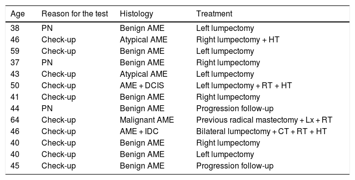
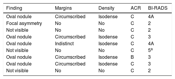
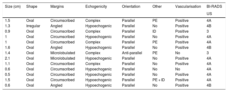
More infoAdenomyoepithelioma of the breast (AB) is an uncommon tumor that is characterized by the biphasic proliferation of epithelial and myoepithelial cells. The radiologic findings for AB have been scantly reported. This paper aims to analyze the characteristic findings for AB on ultrasonography (US) and mammography. This retrospective descriptive study used the Breast Imaging-Reporting and Data System (BI-RADS) to analyze the US and mammography findings for histologically confirmed AB in patients attended at our institution between 2007 and 2019. We identified a total of 13 AB in 13 women. All patients underwent US. On US, 12 lesions were seen as oval nodules, and the remaining lesion was irregular-shaped; 8 lesions had circumscribed margins, 3 had angled margins, and 2 had microlobulated margins. Regarding the echo pattern, 8 lesions were hypoechoic and 5 were complex (solid-cystic). On color Doppler, 11 lesions were vascularized nodules and 2 were avascular nodules. In conclusion, we present the imaging characteristics for a series of cases of AB. It is unlikely that this lesion can be suspected solely on the basis of the imaging findings; nevertheless, it is important for radiologists to be familiar with AB to carry out a good radiopathologic correlation.
El adenomioepitelioma mamario (AME) es un tumor poco frecuente que se caracteriza por la proliferación bifásica de células epiteliales y mioepiteliales. Los hallazgos radiológicos de los AME se encuentran escasamente descritos en la literatura científica. El objetivo de nuestro estudio es evaluar las características de imagen del adenomioepitelioma de mama con un enfoque en la apariencia ecográfica y mamográfica. Se realizó un estudio retrospectivo y descriptivo a partir de las biopsias realizadas en nuestra institución entre 2007 y 2019. Se analizaron retrospectivamente los hallazgos en mamografía y ultrasonido, de acuerdo con el léxico del sistema de informes y datos de imágenes mamarias (BI-RADS), de aquellas lesiones que resultaron con histología positiva para adenomioepitelioma. En el período de estudio se encontraron 13 AME en 13 mujeres. Se realizó ecografía a todas las pacientes; 12 lesiones se presentaron como nódulos ovalados y en un caso con forma irregular. Los márgenes fueron circunscritos (n = 8), angulados (n = 3) o microlobulados (n = 2), el patrón de eco fue hipoecoico (n = 8) y complejo sólido-quístico (n = 5). En cuanto a la vascularización, 11 fueron nódulos vascularizados en Doppler color y 2 resultaron avasculares. En conclusión, presentamos las características imagenológicas de una serie de casos de AME. Es poco probable sospechar esta lesión exclusivamente en función de los hallazgos por imagen; sin embargo, es importante conocer esta lesión para realizar una buena correlación radiopatológica.


