


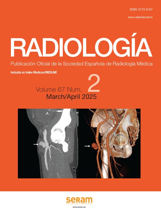


Pseudoaneurysms are common vascular lesions that result as a complication of endovascular interventions, trauma, surgery, inflammatory and tumour processes. They can originate in thoracoabdominal or peripheral visceral arteries. Computed tomography angiography (CT angiography) is the imaging technique of choice for diagnostic confirmation, assessment of complications and therapeutic planning. Doppler ultrasound is initially used in accessible peripheral arteries. The complications of pseudoaneurysms can be potentially serious, mainly their rupture, so their early diagnosis and differentiation from other pathologies is essential to guide treatment. The objective of this manuscript is to represent the image of the pseudoaneurysm in different locations according to its etiology, as well as to review the imaging techniques, study protocols and diagnostic keys. Its complications will be discussed, paying special attention to those that require an emergent therapeutic attitude.
Los pseudoaneurismas son lesiones vasculares frecuentes que resultan como complicación de iatrogenia, traumatismos, procesos inflamatorios y tumorales. Pueden originarse en arterias viscerales toracoabdominales o periféricas. La angiografía por tomografía computarizada (angioTC) es la técnica de imagen de elección para su confirmación diagnóstica, valoración de complicaciones y planificación terapéutica. La ecografía Doppler se utiliza inicialmente en arterias periféricas accesibles. Las complicaciones de los pseudoaneurismas pueden ser potencialmente graves, principalmente su rotura, por lo que su diagnóstico precoz y su diferenciación con otras patologías resulta fundamental para orientar el tratamiento. El objetivo de este manuscrito es representar la imagen del pseudoaneurisma en diferentes localizaciones según su etiología, así como revisar las técnicas de imagen, protocolos de estudio y claves diagnósticas. Se abordarán sus complicaciones, prestando especial atención a aquellas que requieran de una actitud terapéutica emergente.

