metricas
covid
Buscar en
Revista Española de Medicina Nuclear e Imagen Molecular (English Edition)
Toda la web
Inicio Revista Española de Medicina Nuclear e Imagen Molecular (English Edition) Findings of the 18F-FDG PET-CT in a cardiac angiosarcoma complicated by a cardia...
Journal Information
Vol. 33. Issue 4.
Pages 227-230 (July - August 2014)
Share
Share
Download PDF
More article options
Visits
921
Vol. 33. Issue 4.
Pages 227-230 (July - August 2014)
Clinical note
Findings of the 18F-FDG PET-CT in a cardiac angiosarcoma complicated by a cardiac rupture
Hallazgos de la PET-TC con 18F-FDG en un angiosarcoma cardíaco complicado con una rotura cardíaca
Visits
921
Alicia Santiago-Chinchillaa,
Corresponding author
, Eduardo Ruiz-Carazoa, Antonio Moral-Ruizb, Nathalie Testart Dardelb, Alberto Martínez-Martíneza, Silvia López-Fernándezc
a Servicio de Radiodiagnóstico, Hospital Universitario Virgen de las Nieves, Granada, Spain
b Servicio de Medicina Nuclear, Hospital Universitario Virgen de las Nieves, Granada, Spain
c Servicio de Cardiología, Hospital Universitario Virgen de las Nieves, Granada, Spain
This item has received
Article information
Abstract
Full Text
Bibliography
Download PDF
Statistics
Figures (3)
Show moreShow less
Abstract

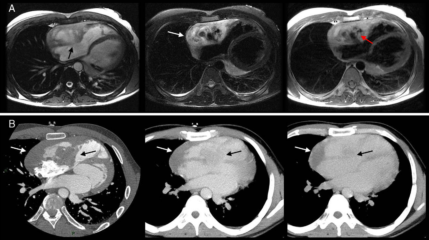
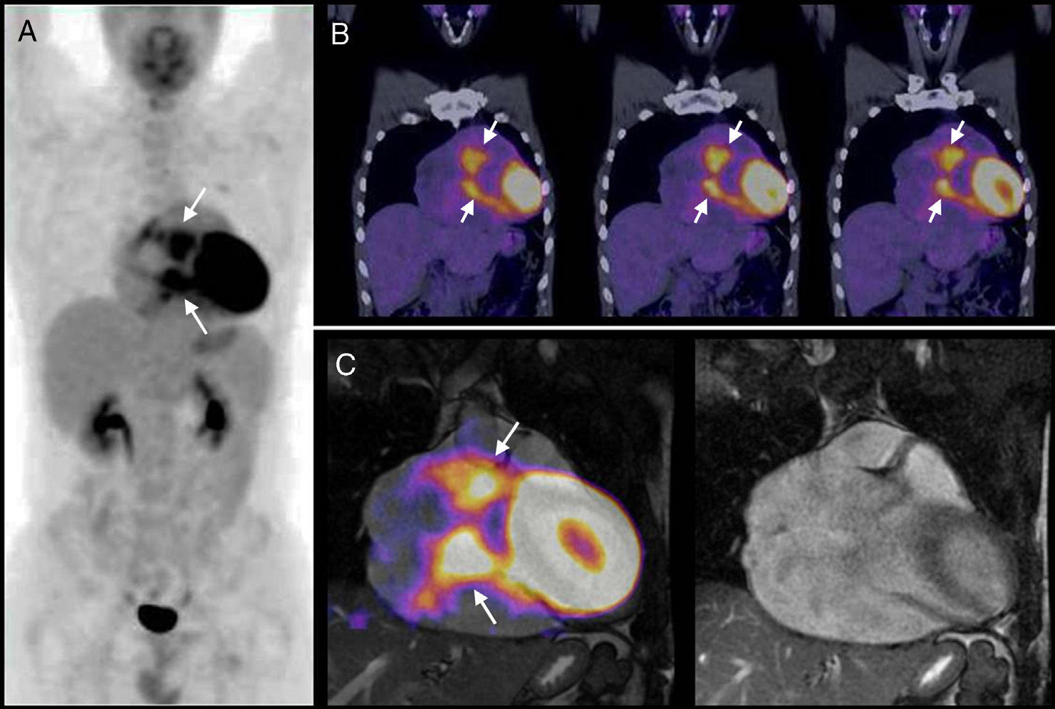
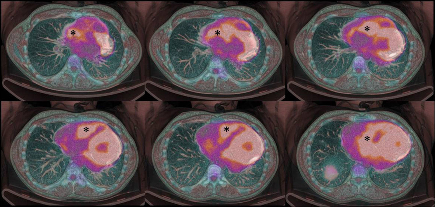
Primary malignant tumors of the heart are a rare condition. The most common type is the cardiac angiosarcoma. The symptoms of this disease are very nonspecific and can be very difficult to diagnose by conventional imaging techniques. We report the case of a male patient with cardiac angiosarcoma who also had a rare complication, this being cardiac rupture, which required the use of 18F-FDG PET-CT to demonstrate the mass malignancy and to reach a definitive diagnosis.

Keywords:
Cardiac angiosarcoma
Computed tomography
Magnetic resonance imaging
18F-FDG
Positron emission tomography-computed tomography
Resumen

Los tumores malignos primarios del corazón son una patología muy poco frecuente y de ellos, el tipo más frecuente es el angiosarcoma cardíaco. Esta patología tiene una clínica muy inespecífica y puede ser muy difícil de diagnosticar por técnicas convencionales de imagen. Presentamos el caso de un paciente con un angiosarcoma cardíaco que además presentaba una complicación muy poco frecuente, una rotura cardíaca, lo que hizo necesario el uso de la 18F-FDG PET-TC para demostrar la malignidad de la masa y poder llegar a un diagnóstico definitivo.

Palabras clave:
Angiosarcoma cardíaco
Tomografía computarizada
Resonancia magnética
18F-FDG
Tomografía por emisión de positrones con tomografía multicorte

Article

These are the options to access the full texts of the publication Revista Española de Medicina Nuclear e Imagen Molecular (English Edition)
Subscriber
Subscriber

If you already have your login data, please click here .

If you have forgotten your password you can you can recover it by clicking here and selecting the option “I have forgotten my password”
Subscribe
Subscribe to

Revista Española de Medicina Nuclear e Imagen Molecular (English Edition)

Purchase
Purchase article

Purchasing article the PDF version will be downloaded

Price 19.34 €

Purchase now
Contact
Phone for subscriptions and reporting of errors
From Monday to Friday from 9 a.m. to 6 p.m. (GMT + 1) except for the months of July and August which will be from 9 a.m. to 3 p.m.
Calls from Spain
932 415 960
Calls from outside Spain
+34 932 415 960
E-mail
Article options
es en pt

¿Es usted profesional sanitario apto para prescribir o dispensar medicamentos?

Are you a health professional able to prescribe or dispense drugs?

Você é um profissional de saúde habilitado a prescrever ou dispensar medicamentos