metricas
covid
Buscar en
Revista Española de Medicina Nuclear e Imagen Molecular
Toda la web
Inicio Revista Española de Medicina Nuclear e Imagen Molecular Valoración preliminar de la 18F-DOPA PET/TC cerebral en el diagnóstico diferen...
Journal Information
Vol. 40. Issue 4.
Pages 214-221 (July - August 2021)
Share
Share
Download PDF
More article options
Visits
2045
Vol. 40. Issue 4.
Pages 214-221 (July - August 2021)
Valoración preliminar de la 18F-DOPA PET/TC cerebral en el diagnóstico diferencial de lesiones cerebrales con RM no concluyente
Visits
2045
B. Domenech Braseroa,
Corresponding author
beatriz.domenech@quironsalud.es

Autor para correspondencia.
, A. Mestre-Fuscob,c, M. Suárez Piñerac, E. Puertas Calvod, X. Perich Alsinae, G. Montesf, P. Plaza Lópeza
a Servicio de Medicina Nuclear, Hospital Quirónsalud Barcelona, Barcelona, España
b Servicio de Medicina Nuclear, Hospital Universitari Doctor Josep Trueta, Girona, España
c Servicio de Medicina Nuclear, Hospital del Mar, Barcelona, España
d Servicio de Oncología Radioterápica, Hospital Quirónsalud Barcelona, Barcelona, España
e Servicio de Radiodiagnóstico, Hospital Quirónsalud Barcelona, Barcelona, España
f Departamento de Neurocirugía, Instituto Clavel, Barcelona, España
This item has received
Disponible módulo formativo: Unidad 4. Saber más
Article information
Abstract
Full Text
Bibliography
Download PDF
Statistics
Figures (2)
Tables (2)
Tabla 1. Características del paciente y tumor, motivo de la prueba PET, tratamiento previo, situación actual y cambio de actitud terapéutica
Tabla 2. Valoración visual y semicuantitativa; SUVmáx, ratio T/S y ratio T/N
Show moreShow less
Resumen
Objetivo

Valorar la utilidad de la 18F-DOPA PET/TC cerebral en el diagnóstico diferencial de lesiones cerebrales con RM no concluyente.
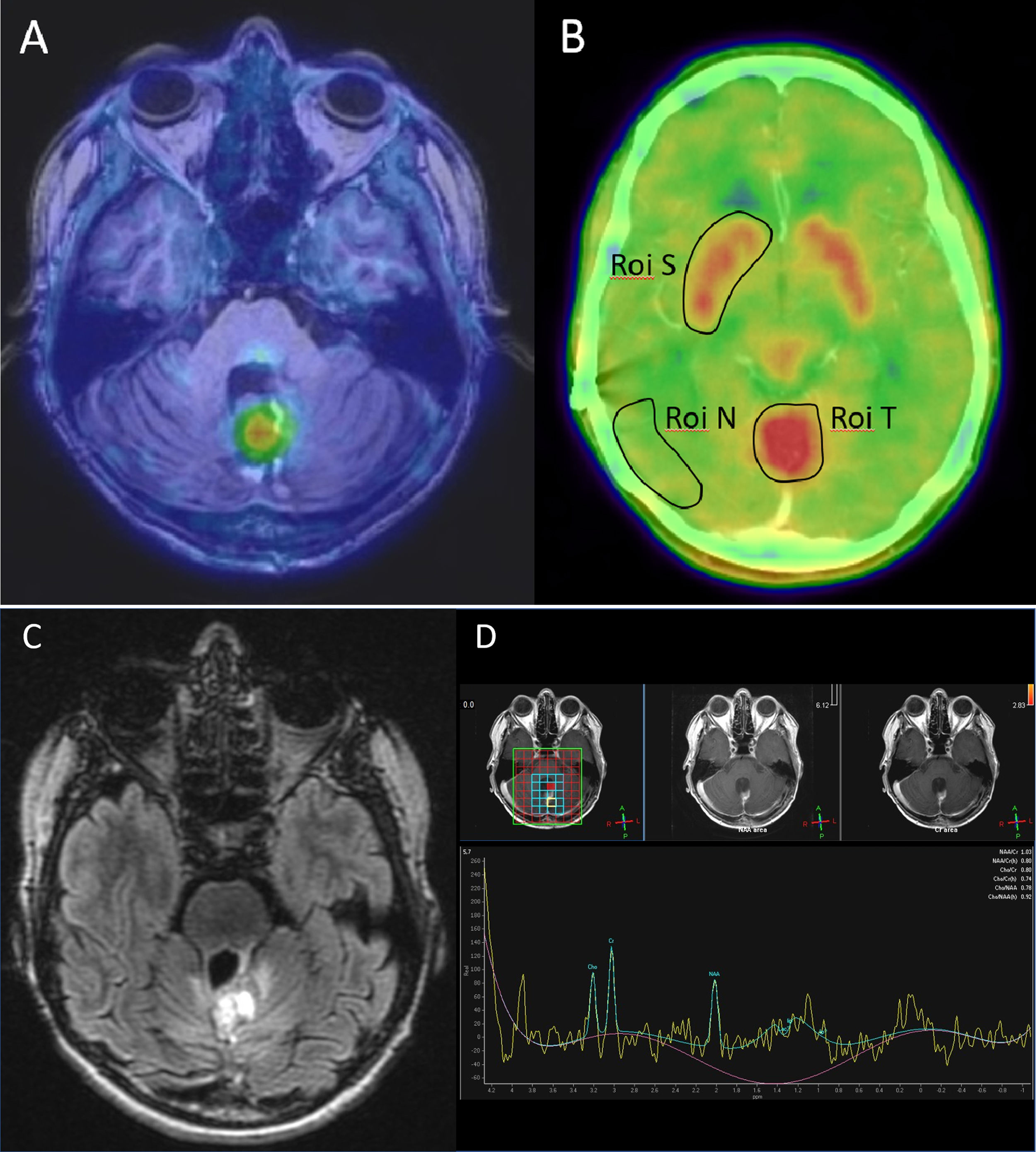
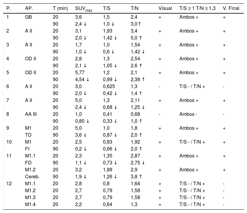
Material y métodos

Se estudiaron 12 pacientes, con un total de 16 lesiones, sin diagnóstico definitivo tras RM cerebral. Se adquirió estudio 18F-DOPA PET/TC cerebral con doble adquisición a los 20 y 90 minutos. Se realizó valoración visual y semicuantitativa con cálculo del SUVmáx de las lesiones y cálculo del ratio T/S (tumor/estriado contralateral) y ratio T/N (tumor/parénquima sano contralateral) para cada uno de los tiempos.
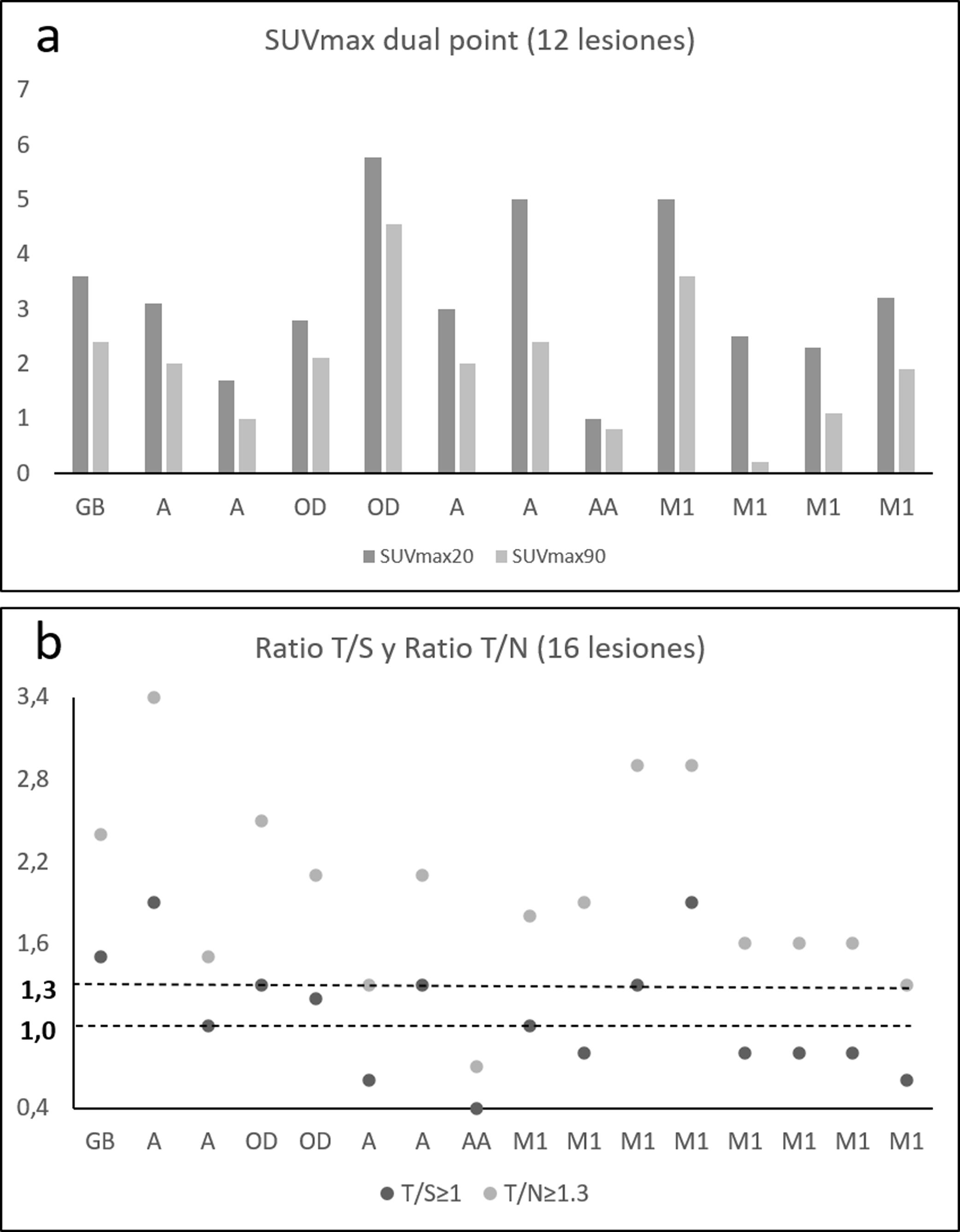
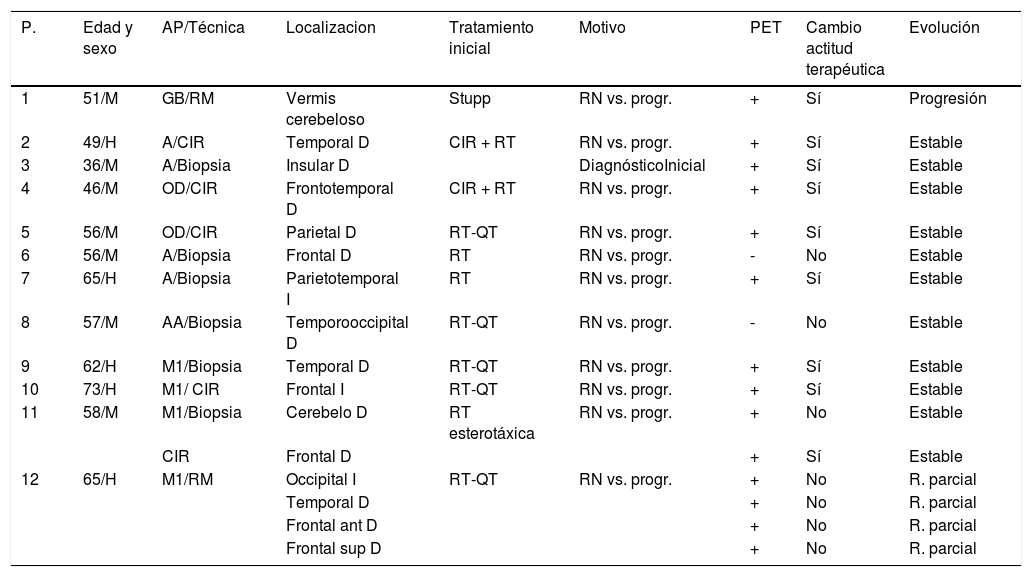
Resultados

Con base en la escala de valoración visual y utilizando los ratios T/S ≥ 1 y T/N ≥ 1,3 para determinar la positividad de un estudio, los valores de sensibilidad (S), especificidad (E) y valor predictivo positivo (VPP) han sido los siguientes: valoración visual (S 100%, E 33,3%, VPP 71,4%), ratio T/S (S 90%, E 100%, VPP 100%) y ratio T/N (S 100%, E 16,6%, VPP 66,6%). Ninguna lesión mostró un aumento del SUVmáx en la adquisición tardía. La 18F- DOPA PET/TC modificó el comportamiento terapéutico en el 75% de los pacientes.

Conclusión

La 18F-DOPA PET/TC es una herramienta útil para diferenciar la etiología de lesiones cerebrales con RM no concluyente. La imagen tardía (dual point) no aporta un valor añadido al diagnóstico final. Los estudios con F-DOPA tienen impacto en el manejo del paciente modificando la conducta terapéutica.

Palabras clave:
PET/TC
18F-DOPA
Glioma
Metástasis cerebral
Recidiva tumoral
Abstract
Aim

To evaluate the utility of brain 18F-DOPA PET/CT in the differential diagnosis of brain lesions with inconclusive MRI.

Material and methods

Twelve patients were studied, with a total of 16 lesions, without definitive diagnosis after brain MRI. A double acquisition PET/CT brain scan was acquired at 20 and 90 minutes. Visual and semiquantitative assessment was performed with SUVmax calculation of the lesions and calculation of the T/S ratio (tumor/contralateral striatum) and T/N ratio (tumor/contralateral healthy parenchyma) for each time.

Results

Based on the visual assessment scale and using T/S ratio ≥ 1 and T/N ratio ≥ 1.3 to determine malignancy, the values of sensitivity (S), specificity (E) and positive predictive value (PPV) were: visual assessment (S 100%, E 33.3%, VPP 71.4%), T/S ratio (S 90%, E 100%, VPP 100%) and T/N ratio (S 100%, E 16.6%, VPP 66.6%). No lesion showed an increase in SUVmax in late acquisition. 18F-DOPA PET/CT modified treatment in 75% of the patients.

Conclusion

18F-DOPA PET/CT is a useful tool in the study of brain lesions with inconclusive MRI. Late imaging (dual-point) has no added value in the final diagnosis. F-DOPA has an impact on patient management modifying therapeutic behavior.

Keywords:
PET/CT
18F-DOPA
Glioma
Brain metastases
Tumoral recurrence

Article

These are the options to access the full texts of the publication Revista Española de Medicina Nuclear e Imagen Molecular
Member
Si es usted socio de la Sociedad Española de Medicina Nuclear e Imagen Molecular (SEMNIM) puede acceder al texto completo de los contenidos de la Revista Española de Medicina Nuclear e Imagen Molecular desde los enlaces a la revista publicados en la web de la SEMNIN (enlace a https://semnim.es/iniciar-sesion/), previo inicio de sesión como socio. Si tiene problemas de acceso puede contactar con la Secretaría Técnica de la SEMNIM en el correo electrónico secretaria.tecnica@semnim.es o en el teléfono: + 34 619 594 780
Subscriber
Subscriber

If you already have your login data, please click here .

If you have forgotten your password you can you can recover it by clicking here and selecting the option “I have forgotten my password”
Subscribe
Subscribe to

Revista Española de Medicina Nuclear e Imagen Molecular

Purchase
Purchase article

Purchasing article the PDF version will be downloaded

Price 19.34 €

Purchase now
Contact
Phone for subscriptions and reporting of errors
From Monday to Friday from 9 a.m. to 6 p.m. (GMT + 1) except for the months of July and August which will be from 9 a.m. to 3 p.m.
Calls from Spain
932 415 960
Calls from outside Spain
+34 932 415 960
E-mail
Article options
es en pt

¿Es usted profesional sanitario apto para prescribir o dispensar medicamentos?

Are you a health professional able to prescribe or dispense drugs?

Você é um profissional de saúde habilitado a prescrever ou dispensar medicamentos