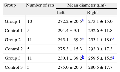
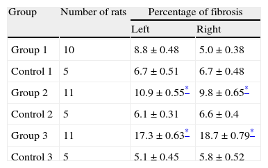
We investigated hypoxia inducible factor-1α (HIF-1α), connective tissue growth factor (CTGF) expression and fibrosis in the testis of rats with surgically induced varicocele.
Materials and methodsA total of 47 adult male Sprague-Dawley rats were arranged in 3 groups, namely group 1 (varicocele operation 4 weeks ago, n=10; sham operation 4 weeks ago, n=5), group 2 (8 weeks, n=11; n=5), and group 3 (12 weeks, n=11; n=5). The rats in every group underwent bilateral orchiectomy 4, 8, and 12 weeks after the operations, respectively. HIF-1α and CTGF expression of both testes in group 3 were studied by real-time reverse transcription-polymerase chain reaction (RT-PCR) and immunohistochemistry. Fibrotic change was assessed by quantitative image analysis.
ResultsHIF-1α mRNA expression in testes tissues in varicocele operation and sham controls showed no significant differences in RT-PCR. However, CTGF mRNA expressions in left testes were found to be significantly different between varicocele operation and sham controls. HIF-1α staining was present in both testes of all specimens and CTGF staining was present in 10 left and 8 right testes of 11 specimens. However HIF-1α and CTGF staining were absent in control group. There were significant fibrotic changes of both testes in groups 2 and 3. There were significant differences in fibrotic change along the durations of surgical varicocele.
ConclusionsThis study reveals that experimental varicocele in the rat is associated with HIF-1α and CTGF expression and it is accompanied by fibrotic change in the testis.
Investigamos la expresión del factor inducible por hipoxia 1α (HIF-1α) del factor de crecimiento del tejido conectivo (CTGF) y la fibrosis en los testículos de ratas con varicoceles inducidos quirúrgicamente.
Material y métodosSe distribuyó un total de 47 ratas Sprague-Dawley adultas en tres grupos: el grupo 1 (4 semanas postcirugía de varicocele, n=10; 4 semanas postcirugía simu-lada, n=5), el grupo 2 (8 semanas, n=11; n=5) y el grupo 3 (12 semanas, n=11; n=5). Todas las ratas de sendos grupos fueron sometidas a una orquiectomía bilateral a las 4, 8 y 12 semanas de la operación respectivamente. Se estudió la expresión del HIF-1α y CTGF de ambos testículos en el grupo 3 mediante RT-PCR e inmunohistoquímica. Se midió los cambios fibróticos mediante análisis cuantitativo de imágenes.
ResultadosLa expresión de HIF-1α mARN en los tejidos testiculares, tanto de la operación de varicocele como de los controles, no mostró diferencias significativas en la RT-PCR. Sin embargo, las expresiones de mARN de CTGF testicular fueron significativamente diferentes entre los de la cirugía de varicocele y los controles. Hubo tinción de HIF-1α en ambos testículos de todos los especímenes y tinción de CTGF en 10 testículos izquierdos y 8 derechos de 11 especímenes. Sin embargo, no hubo tinción de HIF-1α ni de CTGF en el grupo de control. Hubo cambios fibróticos significativos de ambos testículos en los grupos 2 y 3. Hubo diferencias significativas en el cambio fibrótico según la duración del varicocele.
ConclusionesEste estudio revela que el varicocele experimental en la rata se asocia con expresión de HIF-1α y CTGF además de cambios fibróticos en los testículos.
Artículo
Comprando el artículo el PDF del mismo podrá ser descargado
Precio 19,34 €
Comprar ahora


