INTRODUCTION
Pneumomediastinum is an unusual and rare complication of asthma (1-5). During acute attacks of asthma alveolar overdistension and rupture due to excessive air pressure results in the leakage of air from the pulmonary tract and dissection along great vessel sheaths to the mediastinum and pericardium, along the abdominal aorta to the intestinal wall and peritoneum or along fascial planes to the subcutaneous tissue of the neck and axilla. Pneumopericardium is much rare condition air completely surrounds the epicardium and it thus visible in the pericardial sac between the central tendon of the diaphragm and inferior heart border in chest radiographs (1).
In this paper, we describe a 4-year-old girl with asthma who presented with pneumomediastinum, pneumopericardium and subcutaneous emphysema.
CLINICAL CASE
A 4-year-old girl admitted to our hospital with dyspnea, chest pain, palpitation and cough of two days duration. She had experienced attacks of cough, dyspnea and wheezing for two years, but she did not have a diagnosis of asthma previously. Her grandfather had diagnosis of asthma.
Physical examination revealed body temperature of 38 °C, heart rate of 128/min, respiratory rate of 40/min, and a normal blood pressure (100/70 mmHg). She had dyspnea and subcutaneous emphysema in the neck, axilla and thorax. Sibilant rales were heard on auscultation.
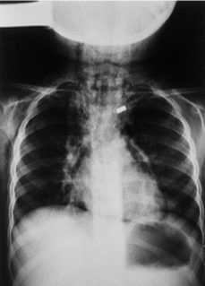
Laboratory findings were following: White blood cell 15,600/mm3 with the predominance of polymorph nuclear leukocytes; eosinophil count: 400/μl; total IgE level: 2,234 IU/l; Phadiatop (Pharmacia, Sweden): positive. In the skin prick test, she had positive reaction to Dermatophagoides pteronyssinus, Dermatophagoides farinae, mold mix, tree mix and grass mix. Pulmonary function test could not be performed. In the chest X-ray air was seen in mediastinum and subcutaneous area and the epicardium was surrounded completely with air (Fig. 1). Her ECG was normal. Two dimensional echocardiography showed mild splitting of visceral and parietal pericardium.
Figure 1.--Chest X-ray of the patient: air was seen in mediastinum and subcutaneous area and the epicardium was surrounded completely with air.
She was treated successfully with nebulised salbutamol and budesonide. Nasal oxygen was administered. Subcutaneous emphysema disappeared in the second day. Radiological signs of pneumopericardium and pneumomediastinum, disappeared completely in ten days period.
DISCUSSION
In recent years, the complications of asthma has been decreased because of the early diagnosis and new treatment regimens. Pneumomediastinum and pneumopericardium are very rare complications of asthma. Incidence of pneumomediastinum has been reported previously as higher as 2-5 % during acute asthma, in a recent report it has been found as 0.3 % (2, 3). These complications results in a worsening clinical course and may be associated with decreased cardiac output and a fall in a blood pressure. Even pneumopericardium developed in our case, she had no sign of pericardial tamponad and cardiovascular deterioration. So she was managed conservatively. Subcutaneous emphysema is a late finding of extra pleural air but it was reported as a most useful predictor of pneumomediastinum (1, 3-5). We also detected subcutaneous emphysema in our case. With the adequate treatment of acute exacerbation further complications of pneumomediastinum and pneumopericardium was prevented.
The time of diagnosis of asthma was another important point in this case. Although she had signs and symptoms of asthma and positive family history she did not have diagnosis of asthma before. We thought that the delayed diagnosis could play a role in the development of these complications.
In conclusion in the light of this case we want to mention that early diagnosis and effective treatment of asthma should be done to prevent serious complication of asthma.


