INTRODUCTION
Eosinophilic esophagitis (EE) is a chronic inflammatory disease of the esophagus characterized by eosinophilic infiltration in the mucosa. An esophageal biopsy is needed to establish the definitive diagnosis. The main symptom is dysphagia, which affects 80 % of all patients, though in children a great variety of symptoms have been described 1-3,5-6.
The underlying etiopathogenesis is presently unknown, though according to most authors an elevated eosinophil count in the esophageal mucosa can be considered an additional manifestation of the allergic disease 5. Previous experimental and in vivo studies have shown an association between EE and allergic disease 1,2,4,8,9. However, we have found no study on the European pediatric population analyzing the associated allergen sensitization pattern.
We carried out a prospective study with 14 children diagnosed of EE in our hospital.
ANALYSIS OF CASES
An analysis was made of the clinical features, which included respiratory allergy, family and personal history of atopia, peripheral eosinophilia and hypersensitivity to inhaled and food allergens, standard blood tests with eosinophil count, serum total IgE and specific IgE to the allergens detected, as well as the conduction of endoscopy, esophageal biopsy and 24-hour pH monitorization. The cutaneous tests were carried out with the prick test, using LETI CBF® for pneumological and food allergens. Fruits and vegetables were also evaluated using prick testing with fresh foods. Serum IgE was determined with Uni-CAP®(Pharmacia).
Of the 14 patients included, 12 were males and 2 females. The mean age was 11 years (range 4-16 years).
The average time elapsed from the onset of symptoms to the moment of diagnosis was 11 months. The clinical features of our patients were similar to those described in previous reports 1,2,5,9. We observed a predominance of males (86 %), and dysphagia and choking as the main symptoms: 71 % of the patients presented dysphagia as the main symptom, associated with choking in 50 %. Other symptoms detected were abdominal pain (28 %), vomiting (14 %), lack of appetite and poor growth (7 %).
In the blood tests, 78 % of the patients showed eosinophilia and 86 % an increase in total IgE. At endoscopy, macroscopic alterations were observed in 5 patients (35 %), 17 % exhibited a whitish pattern in the esophageal mucosa, while the rest of children exhibited "trachealization". The 24-hour monitorization of pH proved normal in all children.
We found a high prevalence of family antecedents of atopia in first degree relatives (50 %), in line with the observations of other reports 1,2,5,9, the main symptom in this sense being allergic rhinitis (41 %).
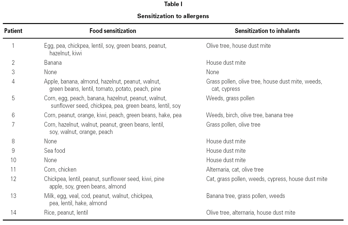
In all patients, prior to the diagnosis of EE, there were symptoms of allergic disease (asthma, allergic rhinitis, food allergy and/or atopic dermatitis) asthma being the most prevalent manifestation (71 %) (table I).
Almost all of our patients presented respiratory allergy in the first phase (asthma, rhinitis), with sensitization to inhalants (93 %), followed by clinical symptoms of EE and sensitization to multiple food allergens though most of them did not have symptoms of food allergy (80 %).
Eleven patients (78 %) showed food sensitization; of these, 8 (73 %) were polysensitized. However, only 2 (20 %) had a history of immediate allergy to food, and none presented anaphylaxis.
In relation to the studies of allergy made, 13 out of 14 (93 %) showed hypersensitivity to aeroallergens, mainly house dust mites (8 patients) and to gramineous species (4 patients), and 50 % showed polysensitization to inhalants. All the patients sensitized to pollens were also sensitized to food. In contrast to the findings of other reports 3,6,10 that point to milk and eggs as the main offenders, the principal form of food sensitization detected in our study was to pulses in 57 % (peanuts, green beans, lentils, green peas, chick peas and soy). Only one of our patients showed sensitization to milk and eggs. Several authors 5,6,9,10 indicate an association of EE to hypersensitivity to aeroallergens and food allergens, which we also observe in our patients. Sensitization associated with EE seems to depend on the alimentary habits of the population studied.
We believe that the high reactivity to legumes in our patients is due to the high intake of such foods in Mediterranean countries 8. A cross-reaction between legumes, and between these and pollens, has been demonstrated by cutaneous and in vitro tests 7,8.
The fact that most of our patients showed sensitization to food and inhalants questions the concept of either alimentary or inhaled allergens. As we can see in the oral allergy syndrome, a mechanism based on primary sensitization to panallergens and the existence of cross-reactivity to inhalants and food is suggested. Respiratory sensitization to panallergens could act as a risk factor for multiple food sensitization and for the posterior onset of EE.
We conclude that further studies are needed in patients with respiratory allergy in order to draw firm conclusions.
Correspondence:
Ana Maria Plaza Martin
Hospital Sant Joan de Déu
Servicio de Alergología
P.º Sant Joan de Déu, 2
08950 Esplugues. Spain
E-mail: aplaza@hsjdbcn.org


