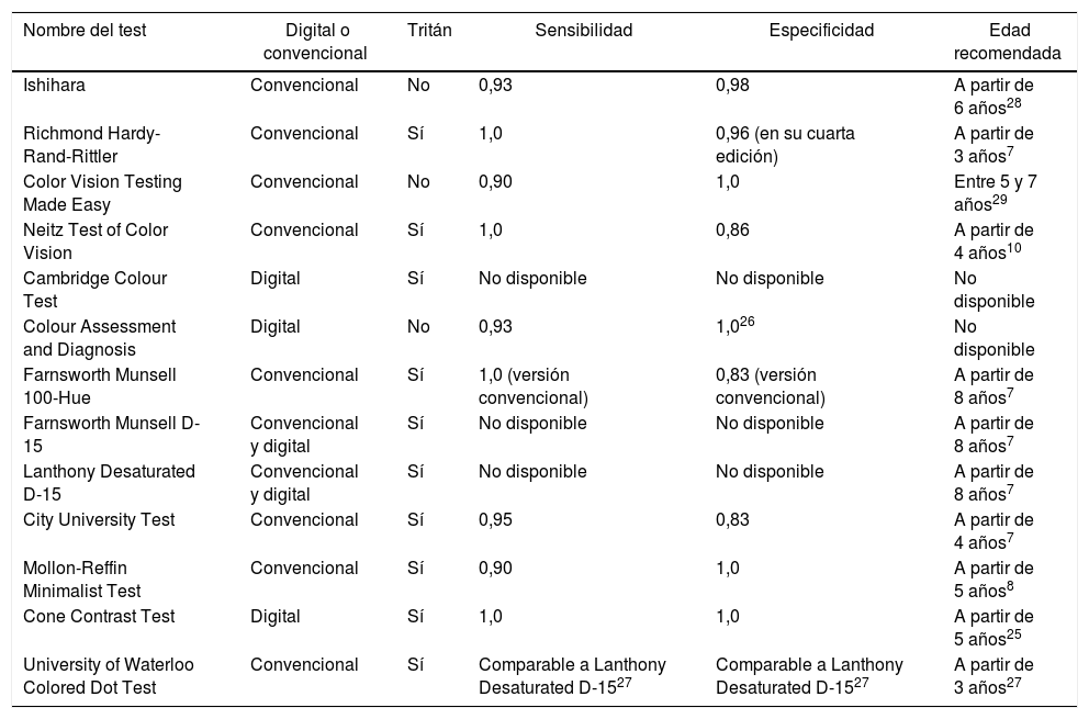
Las deficiencias congénitas en la visión del color afectan a un 8% de la población masculina y a un 0,5% de la femenina. El estudio de la visión del color es un proceso complejo debido a diversos factores: la propia psicofísica de la visión y la dificultad de establecer modelos matemáticos para su análisis, la vaga correlación de los resultados entre unos test y otros y la influencia de factores externos como la iluminación, la condición de los test o la experiencia del examinador y del paciente. En el presente documento se realiza una revisión simplificada de los principales test disponibles en la práctica clínica para evaluar la visión del color.
Material y métodosTras realizar una filtrada revisión preliminar de la bibliografía relacionada con el estudio de la visión del color en el motor de búsqueda PubMed, se determinaron los test mayormente utilizados en la práctica clínica. Se realizó una interpretación atendiendo a su frecuencia de uso y el propósito para el que eran utilizados. A continuación, se procedió con un estudio bibliográfico de cada test en particular, atendiendo al diseño de los estímulos presentados, su población diana y su sensibilidad y especificidad.
ResultadosDe las 95 publicaciones que mostró el buscador PubMed, en 41 de ellas los investigadores utilizaron test de colores en su metodología. De los 64 test de color utilizados, 19 eran diferentes (contando como distintos los test adaptados por grupos de investigación, 4, y aquellos realizados online, 2). El orden de empleo de los test es el siguiente: test de Ishihara (10,88%), Farnsworth-Munsell (7,04%), Farnsworth-Munsell 100 Hue (6,4%), Cambridge Colour Test (3,84%), Hardy-Rand-Rittler (3,2%), test propios desarrollados por los grupos (2,56%), el anomaloscopio (1,28%), los test online (1,28%) y, finalmente, Colour Assessment and Diagnosis (0,64%), Pflüger Trident Colour Plates (0,64%), Toothguide Training Box (0,64%), Lanthony Desaturated D-15 (0,64%), City University Test (0,64%), Universal Colour Discrimination Test (0,64%) y Rabin Cone Contrast Test (0,64%).
ConclusionesEl gold standard en cuanto a la evaluación de la visión del color es el anomaloscopio, instrumento incompatible con la práctica clínica diaria. Su manejo es relativamente complicado, exige disponibilidad de tiempo para su aplicación y es difícilmente comprensible por población infantil. Sin embargo, es posible alcanzar una fiel aproximación mediante la combinación de algunos de los test enumerados en este artículo. Los test expuestos son una buena alternativa para determinar la presencia de discromatopsias en ambientes cercanos a la práctica clínica diaria o en entornos menos controlados que un estudio clínico. El inconveniente principal del amplio elenco de test disponibles para el estudio de la visión del color es la dificultad para comparar los resultados entre test, ya que los datos publicados suelen tener unidades distintas, requiriendo experiencia para su correcta interpretación. En la actualidad, no existe unanimidad sobre qué test de color resulta ser el más completo; es recomendable utilizar al menos 2 para asegurar los diagnósticos y tener una información más completa sobre la percepción visual de los pacientes.
Congenital colour vision deficiencies affect 8% of the male and 0.5% of the female population. The study of colour vision is a complex process due to several factors: the psychophysics of vision itself, the difficulty to establish mathematical models for its analysis, the vague correlation of results between different tests, and the influence of external factors such as lighting, the tests condition, or the experience of the examiner and the patient. In the present document, a simplified review was carried out on the main colour vision tests available in clinical practice.
Material and methodsOnce a filtered preliminary review was made of the bibliography related to the study of colour vision using the PubMed search tool, the most used tests in clinical practice were selected according to their frequency of use and the purpose for which they were applied. A bibliographic study was then carried out on each particular test according to the design of the shown stimuli, its target population, and its sensitivity and specificity.
ResultsFrom the 95 publications found using the PubMed search tool, in 41 of them, colour tests were used by researchers in their methodology. From the 64 colour tests used, 19 of them were different (with 4 of them being different tests adapted by research groups, and 2 of them carried out online).
The most used tests were the following: Ishihara test (10.88%), Farnsworth-Munsell (7.04%), Farnsworth-Munsell 100 Hue (6.4%), Cambridge Colour Test (3.84%), Hardy-Rand-Rittler (3.2%), tests developed by the groups (2.56%), the Anomaloscope (1.28%), the online tests (1.28%) and, finally, Colour Assessment and Diagnosis (0.64%), Pflüger Trident Colour Plates (0.64%), Toothguide Training Box (0.64%), Lanthony Desaturated D-15 (0.64%), City University Test (0.64%), Universal Colour Discrimination Test (0.64%), and Rabin Cone Contrast Test (0.64%).
ConclusionsThe Anomaloscope is the “gold standard” in terms of colour vision testing, despite its incompatibility with daily clinical practice. It is fairly complex to use, difficult to understand for children, and its practice requires having the time available. Nevertheless, it is possible to reach an accurate approximation through the combination of some of the tests listed in this article. The above mentioned tests are a good alternative to determine the presence of dyschromatopsia in settings closer to daily clinical practice or in less controlled settings than a clinical study. The major drawback among the wide range of tests available for the study of colour vision is the difficulty to compare results between tests, since units of the reported data are usually different, and experience is required for its correct interpretation. Currently, there is no consensus on which colour test is the most complete. It is, therefore, advisable to use at least 2 tests in order to ensure diagnoses, and have more extensive information about the visual perception of patients.
Artículo
Comprando el artículo el PDF del mismo podrá ser descargado
Precio 19,34 €
Comprar ahora


