Evaluar diferentes parámetros del segmento anterior en ojos con pseudoexfoliación (PSX), ojos contralaterales y controles mediante tomografía de coherencia óptica (OCT) y cámara Scheimpflug.
MétodosSe estudiaron 3 grupos: 44 ojos de 44 pacientes con PSX, 30 ojos contralaterales no afectos y 148 ojos de 148 controles sanos. Mediante la cámara de Scheimpflug (Pentacam, Oculus Inc.; Wetzlar, Alemania) se midieron la profundidad y volumen de la cámara anterior, volumen corneal y paquimetría, diámetro pupilar y densitometría corneal. Mediante OCT RTVue 100 (Optovue, Fremont, CA, EE. UU.) se midieron la abertura angular, la longitud y el área de la malla trabecular, el grosor del iris, y se valoró la visualización de depósitos PSX.
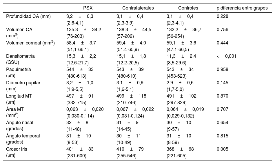
ResultadosNo se observaron diferencias en cuanto a la profundidad ni volumen de la cámara anterior, ni en el volumen corneal o paquimetría (p≥0,228 en todos los parámetros) entre grupos. Sin embargo, la densitometría corneal fue mayor en PSX y en los ojos contralaterales que en el grupo control (p<0,001). En cuanto a los parámetros de OCT no existieron diferencias en la abertura angular ni en el tamaño de la malla entre los 3 grupos, siendo el grosor del iris menor en controles (p=0,005); identificándose en todos los pacientes el depósito PSX mediante OCT.
ConclusionesNo se detectaron diferencias entre las medidas biométricas del segmento anterior entre los pacientes con PSX y controles, salvo en el caso de la densitometría corneal central y el grosor del iris que fueron mayores en el grupo con PSX y en los ojos contralaterales.
To evaluate different anterior segment parameters in eyes with pseudoexfoliation (PSX), fellow eyes, and controls using optical coherence tomography and a Scheimpflug imaging system.
MethodsThree groups were studied: 44 eyes of 44 patients with PSX, 30 clinically unaffected fellow eyes, and 148 eyes of 148 healthy controls. The anterior chamber depth and volume, corneal volume and thickness, pupil diameter and corneal densitometry were measured using a Scheimpflug imaging system (Pentacam, Oculus Inc.; Wetzlar, Germany). The angle width, the length and area of the trabecular meshwork, and the iris thickness were measured using an optical coherence tomography RTVue 100 device (Optovue, Fremont, CA, USA). The presence of PSX deposits was also assessed by OCT.
ResultsThere were no differences in the anterior chamber volume or depth in the corneal volume or central thickness (P≥.228). The corneal densitometry was similar between PSX and fellow eyes; however it was greater than in the control group (P<.001). As regards the parameters measured by OCT, there were no differences in the angle width or in the trabecular meshwork size between the 3 groups; however, the iris was thinner in controls (P=.005). In all patients the PSX deposits were correctly visualised by OCT after the identification by biomicroscopy.
ConclusionsThere were no differences in the anterior segment biometric measurements between patients with PSX and controls, although the corneal densitometry and iris thickness were greater in the PSX and fellow eyes groups.


