Desarrollar modelos predictivos de morfometría corneal en cirugía refractiva con láser excímer para la corrección de ametropías.
MétodoSe realizó una investigación longitudinal y prospectiva con 78 pacientes (151 ojos) operados con LASIK y 56 pacientes (111 ojos) operados con LASEK. Se utilizó el microscopio confocal ConfoScan 4 de NIDEK. Se aplicó el ANOVA de un factor con corrección de Bonferroni, correlación de Pearson y análisis de regresión lineal múltiple con validación cruzada.
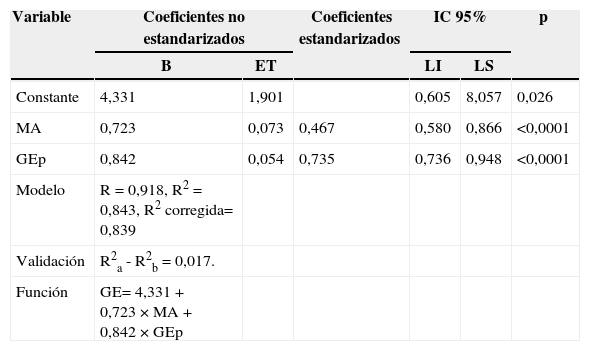
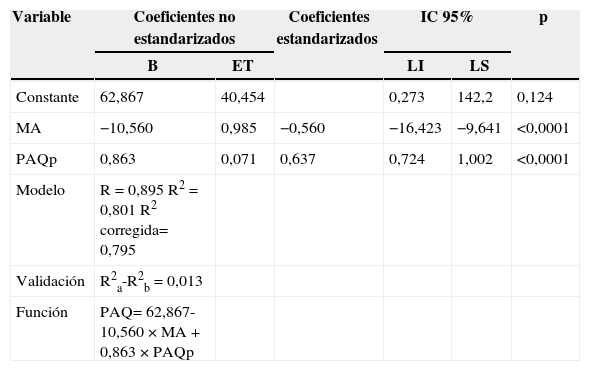
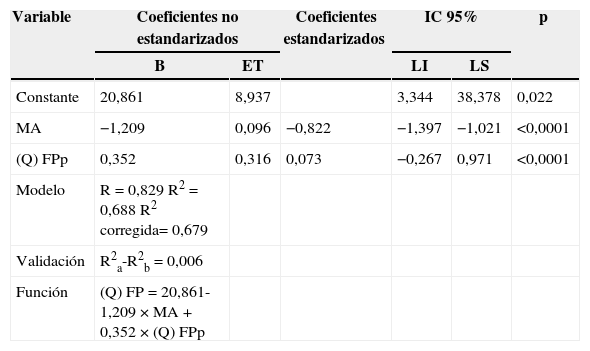
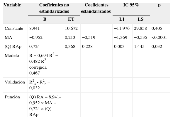
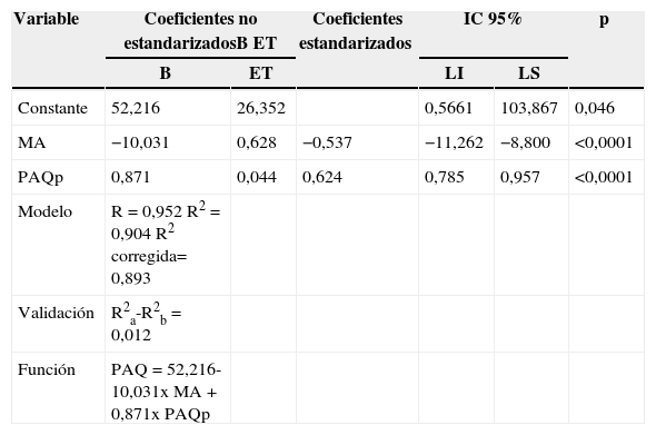
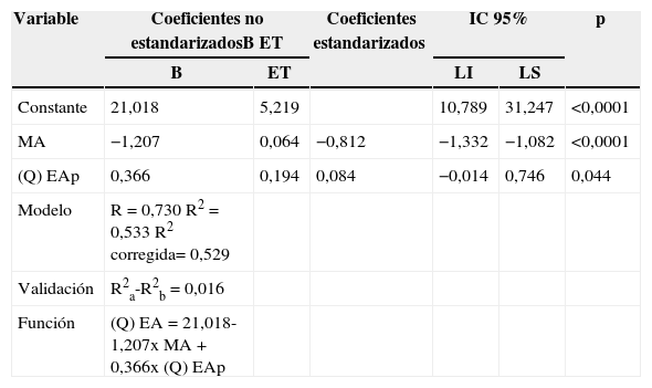
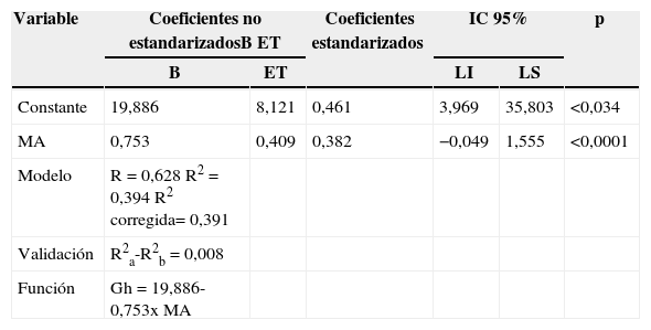
ResultadosTras el LASIK, el 84,3% de las variaciones del grosor epitelial se deben a la magnitud de la ametropía tratada y al grosor epitelial preoperatorio. El 68,8 y el 48,2% de las variaciones de la densidad de queratocitos en el colgajo posterior y zona de retroablación anterior, respectivamente, se deben a los valores de estas variables antes de LASIK y a la magnitud de la ametropía tratada. Tras el LASEK el 90 y el 53% de las variaciones de paquimetría corneal y densidad de queratocitos al año, respectivamente, se deben al valor de esta variable en el preoperatorio y a la magnitud de la ametropía tratada.
ConclusionesLos modelos predictivos obtenidos revelan que las variaciones de las variables morfométricas al año del tratamiento dependen en gran medida de sus valores preoperatorios y de la magnitud de la ametropía a tratar. Estos modelos constituyen herramientas a tener en cuenta como criterios de selección de pacientes candidatos a cirugía con láser excímer para tratamiento de ametropías, y para la elección óptima de la técnica quirúrgica.
To develop corneal morphometric models with refractive error in excimer laser surgery.
MethodA prospective-longitudinal study was conducted on 78 patients (151 eyes) using the LASIK surgical technique, and 56 patients (111 eyes) with myopic astigmatism using ESIRIS (Schwind-Germany) equipment with pendulous microkeratome. The results were analyzed using descriptive statistics. A NIDEK Confoscan microscope was used to obtain and study the images.
ResultsAfter LASIK treatment 84.3% of the variations in epithelium thickness variations were due to the magnitude of refractive error and the epithelium thickness before LASIK treatment. More than two-thirds (68.8%) of the variations in keratocyte density variations in posterior flap and 48.2% of the variations in the anterior retroablation zone were due to the magnitude of the refractive error. Variations of 90% were found in the corneal thickness after LASEK, which were due to the magnitude of the refractive error before LASEK.
ConclusionsPredictive models reveal that morphometrical variations depend of the magnitude of the refractive error. These models are very important in the selection of patient for refractive surgery, and also for the specific technique to use.
Artículo
Comprando el artículo el PDF del mismo podrá ser descargado
Precio 19,34 €
Comprar ahora


