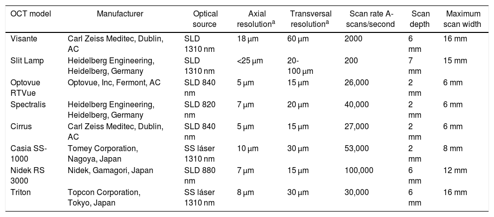
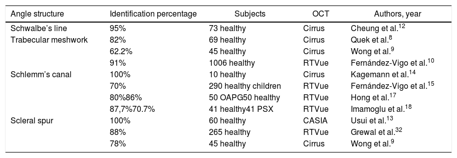
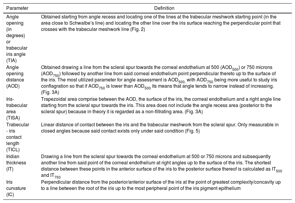
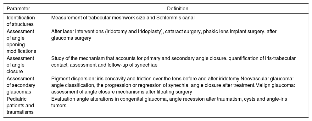
The iridocorneal angle, due to its implications in the physiopathology of aqueous humour drainage, is a fundamental structure of the anterior chamber. Anterior segment optical coherence tomography (AS−OCT) is a rapid and non-invasive technique that obtains images in vivo. The high resolution allows it to analyse the normal anatomy of the angle, any alterations, and the changes that occur after different therapeutic interventions. AS−OCT technology has evolved to provide images that allow the identification and quantification of the angular structures in healthy subjects and in glaucoma patients, and especially the trabecular meshwork and the Schlemm's canal. It also enables the angle width to be quantified, with some objective parameters that have been standardised in recent years, such as the trabecular-iris angle (TIA), the angle opening distance (AOD), and the trabecular-iris area (TISA). This technique has multiple uses in the study of the different mechanisms of angle closure, the evaluation of changes after a laser peripheral iridotomy or iridoplasty after cataract surgery, as well as after the implantation of phakic lenses.
El ángulo iridocorneal por sus implicaciones en la fisiopatología del drenaje del humor acuoso es una estructura fundamental de la cámara anterior. La tomografía de coherencia óptica de segmento anterior (OCT-SA) es una técnica rápida y no invasiva que obtiene imágenes de los tejidos vivos con una alta resolución permitiendo conocer la anatomía normal del ángulo, sus alteraciones y los cambios que se producen en el mismo tras diferentes intervenciones terapéuticas. La tecnología de la OCT-SA ha ido evolucionando hasta ofrecer imágenes que permiten identificar y cuantificar estructuras angulares claves en sujetos sanos y en pacientes con glaucoma, especialmente la malla trabecular y el canal de Schlemm, lo que puede contribuir a ampliar el conocimiento de la fisiopatología del glaucoma. Además, permite cuantificar la abertura angular con unos parámetros objetivos descritos en los últimos años, entre los que destacan el ángulo irido-trabecular (TIA), la distancia de abertura angular (AOD) y el área irido-trabecular (TISA). La OCT-SA presenta múltiples utilidades en el estudio de los distintos mecanismos del cierre angular, la evaluación de los cambios angulares tras la realización de una iridotomía láser o iridoplastia, cirugía de la catarata o el implante de lentes fáquicas.
Artículo
Comprando el artículo el PDF del mismo podrá ser descargado
Precio 19,34 €
Comprar ahora


