Introduction
Diabetic retinopathy (DR) is a highly prevalent, chronic and progressive disease. It is the second cause of blindness in Spain and the second in people of working age.1 Even in its most aggressive forms, loss of acute vision symptoms are not normally present, so when there is a decrease it is usually too late for effective treatment. For this reason early detection is vital.
Laser photocoagulation prevents or delays vision loss in a good number of patients.2 Photographs of the back of the eye are more reliable than ophthalmoscope for diagnosis.3 Studies have validated the digital image as the most ideal method for DR screening.4
In the Andalusian Health System the diagnosis and follow-up of type 2 diabetic patients is carried out in primary care centres. In those centres, the health network allows digital images taken, to be sent by e-mail to a reference centre to be stored and studied by ophthalmologists for classification and treatment of DR.
Studies are required to assess the real use of this method and, its practicality in providing full cover for the diabetic population, and its direct relationship at the 2 care levels and, its potential to reduce costs with a greater benefit for patients and the system.
To achieve this, we proposed the following objectives:
to evaluate the concordance of biomicroscopy between ophthalmologists. To analyse the validity of digital photographs (received by e-mail) read by ophthalmologists and family doctors to detect DR, and to look at the benefits for patients.
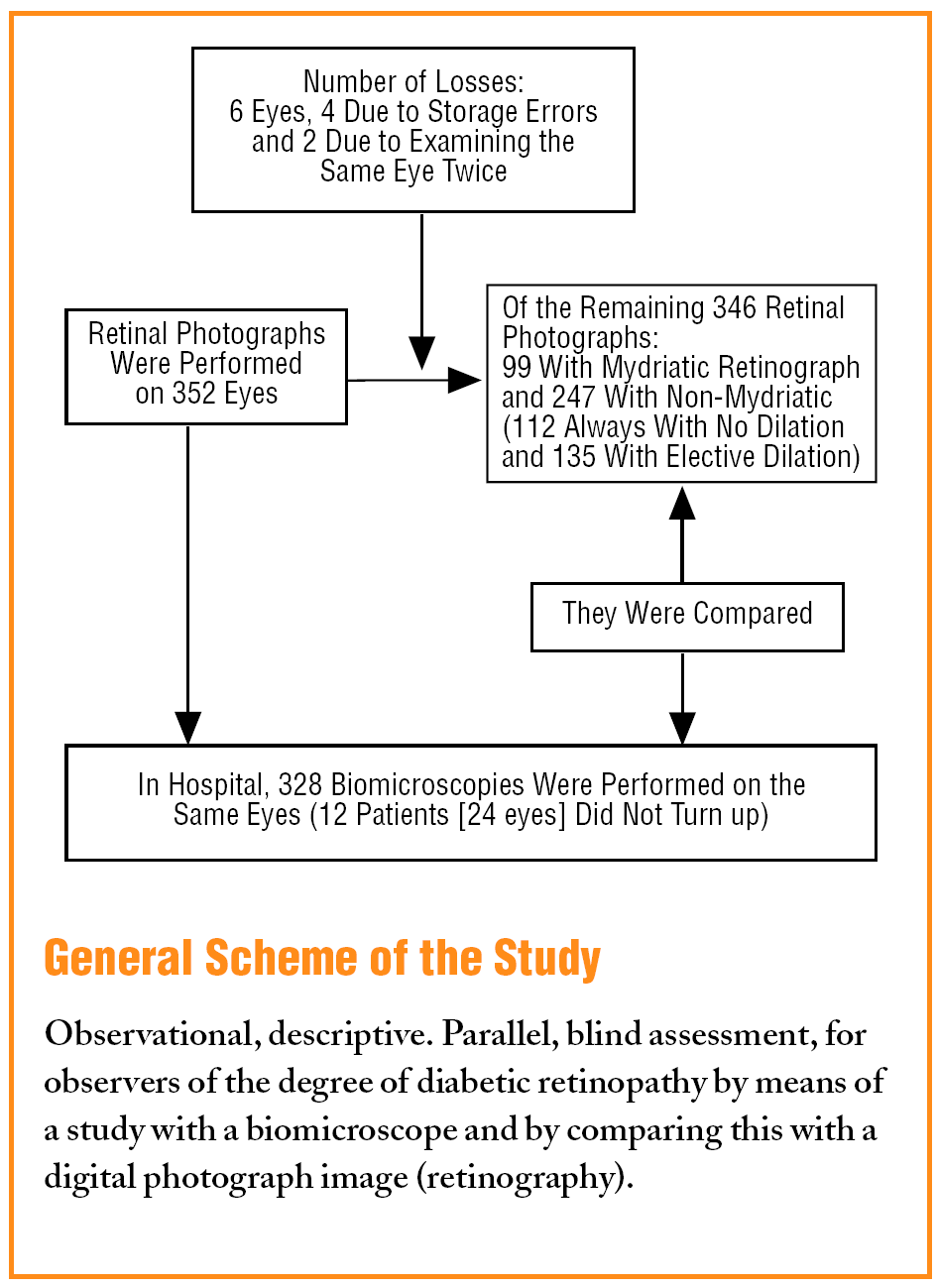
General Scheme of the Study. Observational, descriptive. Parallel, blind assessment, for observers of the degree of diabetic retinopathy by means of a study with a biomicroscope and by comparing this with a digital photograph image (retinography).
Methods
Design
Observational, descriptive. Parallel blind assessment, of the degree of DR measured with a biomicroscope and comparing this with a digital photographic image.
Population and Sample
Type 2 diabetic patients, who had not received photocoagulation, consecutively selected on arriving at a clinic.
The sample size for the kappa index is N=196 (15% disagreement ratio, precision 5%, and a 95% confidence level).5 For validation, for a 95% negative predictive value, precision 5% and a 95% confidence level, N=73.
It was increased to Na=91, according to Na=N [1/(1R)], for losses.6
Measured Variables
Degree of DR in the digital photograph and biomicroscopy, according to the modified Early Treatment Diabetic Research Study (ETDRS) classification,7 measured by an ophthalmologist. Macular oedema was excluded as stereoscopic photographs could not be taken.
Diagnosis of DR in the photograph by the family doctor.
Data Collection and Variable Measurement Techniques
In a first group of participants, a family doctor, previously trained in ophthalmology, took 3 photographs (JPEG format) of each eye with a Topcon® TRC-50 EX mydriatic retinograph, under mydriasis with tropicamide with or without phenylephrine, and of the, macular, nasal and upper temporal fields.8 With the second group, a Topcon® NW100 non-mydriatic retinograph was used, with first ones always without dilation and the following ones dilated with tropicamide when, in their opinion, the quality of the photograph was not good. In both cases, 3 photographs, nasal, central, and temporal (immovable fixed points in this retinograph), were taken.
The photographs were sent by e-mail to the Juan Ramón Jiménez Hospital, using the program Outlook 2000®, over the Andalusia Regional Government Corporate network.
The eye fundus (3 photos) was divided into 6 fields: papilla, macula, upper, lower, temporal, and nasal; it was considered "not assessable" when more than 3 were of poor quality.
The patients were sent to the ophthalmologist with 3 weeks to avoid any progression of the DR causing discordances.
Biomicroscopy was performed independently by 2 ophthalmologists with a slit lamp and a non-contact lens (VOLK Super 66)® or a contact lens (Ocular MAINSTER Standard Focal/Grid®), if the version was inappropriate.
Data Analysis
Weighted linear and quadratic kappa index, was used to assess the agreement of the biomicroscopy between ophthalmologists. For the strength of the agreement the Landis and Koch9 scale was used. To test the validity, sensitivity (Se), specificity (Sp), the positive predictive value (PPV), and the negative predictive value (NPV), for all the standards and confidence intervals (CI) were measured.
Version 11.0 of the SPSS program and version 6.0 of EpiInfo program was used.
Results
There were 352 retinal photographs performed; 99 with a mydriatic retinograph (MYD), 247 non-mydriatic (NOMYD): 112 always with no dilation (NOMYD-ND) and 135 with elective dilation (NOMYD-D). Six eyes (1.7%) eyes were lost, 4 due to storage errors and 2 due to duplication of the examination in the same eye. There were no losses in the electronic transmission.
The mean age and standard deviation of the patients was 65.4±9.9 years.
The ophthalmologist considered 28% of the examinations non-assessable (17.2% of those performed with MYD, 38.4% of the NOMYD-ND, and 27.4% of the NOMYD-D) (P=.0027). Cataracts were more common in the non-assessables (63.42%) than in the assessables (15.32%) (P<.001).
Similarly with other problems of transparency (5.8% compared to 4.9%; P=.005). A family doctor made 291 examinations, of which 23.71% were non-assessable compared with 50.9% which made 55 (P=.004).
Of the retinal photographs read by ophthalmologists, 25.7% had DR: 10.4% mild (MiDR), 12% moderate (MoDR), and 3.2% severe (SDR).
The family doctor considered 13.6% of the retinal photographs non-assessable. Of the assessable ones, 36.5% had DR.
Overall, 328 biomicroscopies were performed, as 12 of the patients (24 eyes) did not turn up, 3 of them had lesions in both eyes, and in 1 the lesion was MoDR. Four were non-assessable (1 due to a detached retina, 2 due to cataracts, and 1 with no diagnosis). Of the 28.7% who had a lesion: 12.65% were MiDR; 13.9%, MoDR; 1.8%, SDR; and 0.3% very severe (VSDR). The mean time between the retinograph and the biomicroscopy was 15.6 days, and ranged between 2 and 68 days.
The agreement in biomicroscopy between ophthalmologist was "very good" (linear weighted kappa =0.80; 95% CI, 0.73-0.88, and quadratic weighted kappa =0.88; 95% CI, 0.65-0.95).
Table 1 shows the validity results of digital photographs read by the ophthalmologists and family doctors as a method for detecting DR and referable DR (grade ≥MoDR), and in Table 2, that of the ophthalmologists differentiated by retinal photographs. Significant differences were found in sensitivity in favour of MYD (P=.009), compared to NOMYD-ND.
Discussion
The agreement in biomicroscopy was "very good." It enables the examination by an ophthalmologist to be like a reference test.
The time between the 2 tests was not seen to influence the differences, since it was too short for the lesions to progress.
We found the sensitivity to be slightly less than the 80% recommended by the British Diabetic Association10 and a specificity slightly higher than the recommended 95%. With NOMYD-SD, the sensitivity is clearly lower, although specificity is maintained. This decrease in sensitivity is found by the majority of authors. When the eyes which had had poor quality photographs were subsequently dilated, we found a great improvement in the sensitivity, although lower than MYD dilating to 100% and with higher temporal field. In this case, the sensitivity and specificity are similar to those of Baeza Díaz et al,11 using 3 fields and dilation if the examination quality was poor, or Murgatroyd et al,12 who used 3 fields with dilation, and somewhat higher than that of other authors such as Harding et al13 and Lawrence,14 with 3 fields, or Scanlon et al,15,16 Olson et al17, or Stellingwerf et al,18 with 2.
The negative predictive value was taken as a reference for the sample size, since, on having an effective treatment for DR, it is important to avoid the patients with a lesion being diagnosed as healthy. In our case it was very good and similar to that found by Stellingwerf et al18 and lower than that of Baeza Díaz et al.11
Although 28% of the examinations were non-assessable, it would be 5.8% if we discard those who had cataracts or opacities, which is around the 5% recommended by the British Diabetic Association.10 If we analyse by methods, the losses decrease with dilation, and this difference is statistically significant. Murgatroyd et al,12 achieved a reduction in non-assessables from 26% to 5% by dilating the eyes to 100%. The difference found between the MYD and NOMYD could be due to 3 different causes: the first could be the dilation of the eyes to 100% instead of 50.74% and, also a higher dilation by using 2 drops of tropicamide and phenylephrine if 1 was not sufficient. The second could be the greater temporal field which cannot be done with NOMYD. This would have less influence because, although some authors, such as Baeza et al,11 found slight improvements in the sensitivity for retinopathy, others, such as Perrier et al19 and Baeza et al themselves, did not find any when they studied referable retinopathy. Also, the third field does not reduce the losses. The third reason could be the difference in technical quality of the retinal photographs. That from the MYD was higher than the NOMYD used, which was a portable apparatus with poorer software.
On the other hand, many disadvantages were observed: MYD required much more time to learn than NOMYD and it resulted in one of the 3 doctors refusing to use it; the time required, 12 examinations were made with NOMYD for every 5 with MYD, with the need for more health staff, and lastly, the inconvenience for the patients, longer waiting time in clinic and then a much longer and uncomfortable pupil dilation.
The reading of the digital photograph by a family doctor is very safe when detecting a significant DR. Good sensitivity was found, 95.2%, and a very good NPV of 99% when MoDR or higher was assessed, which ensures that patients with DR are not diagnosed as healthy. However, the specificity of 81.5% and a PPV of 47.6% to detect MoDR or higher, raises doubts on if the high number of false positives in a disease like DR, which could mean telling patients that they do not have it, counteracts the benefits that practically no lesion escaped. In any case, it is clear that training in reading, the only objective difference between these and the ophthalmologist, is essential.
As regards benefits to the patients (and for the health system), half of them would have avoided going to the ophthalmologist and almost all of them that had to go had a DR (16.8%≥MoDR) or opacities.
To send the digital photograph of the back of the eye of type 2 diabetics by e-mail, as a method of detecting and monitoring DR, is viable and on setting it up the following recommendations should be taken into account:
The retinal photograph should be the non-mydriatic type, since the management of mydriatic is too complex and slow
The retinal photograph should be taken by photographing at least 2 retinal fields of 45° (a single field has been rejected by several authors due to its insufficient validity,11,12,14,16,20,21 and dilating with tropicamide, as it significantly improves sensitivity and the percentage of assessable photographs
If the family doctor is going to read the photographs in the health centre, training must be improved to avoid the high number of false positives found
The person who performs the retinal photographs should do the highest number possible, since the percentage of non-assessables significantly decreases with practice
With these recommendations, it would mean that only between 30% and 40 % of patients would have to be seen by the ophthalmologist in the first visit, and given the high percentage of opacities in the non-assessables, it is likely that in the following reviews the percentage of referrals would be even less on this problem being controlled.
Project financed by the Health Research Fund (FIS 01/0708) of the Ministry of Health, which made the acquisition of the retinal photographs possible.
The project received assistance from the Andalusian Society of Family and Community Medicine.
What Is Known About the Subject
• Diabetic retinopathy is highly prevalent disease which does not usually present with loss of acute vision; its early detection is essential.
• Back of the eye photographs are more reliable than ophthalmoscope for the diagnosis of diabetic retinopathy.
• The health service internet allows digital images taken in primary care health centres to be sent, stored and studied by ophthalmologists.
What This Study Contributes
• Retinal photographs of type 2 diabetic patients sent by e-mail is feasible as a method for the detection and monitoring of retinopathy.
• The retinograph must be the non-mydriatic type and the photos taken dilated with tropicamide.
• The person taking the retinal photographs should do as many as possible and be trained well to avoid losses and the high number of false positives.
Correspondence:
Dr. E. Molina Fernández.
Unidad Docente de Medicina.
Familiar y Comunitaria de Huelva.
Servicio Andaluz de Salud.
Hospital Vázquez Díaz. 4.a planta.
Ronda Norte, s/n. 21005 Huelva.
España.
E-mail: udhuelva@tiscali.es
Manuscript received February 26, 2007.
Manuscript accepted for publication October 8, 2007.
Spanish version available
www.doyma.es/197.924


