


Urachal sinus (US) is a rare congenital abnormality resulting from the improper involution of embryological tissues connecting the bladder to the umbilicus. It typically presents in childhood but can remain asymptomatic into adulthood.
A 70-year-old male with hypertension and dyslipidemia presented with periumbilical pain, swelling, and purulent discharge. Initial empirical treatment with antibiotics was ineffective. Further imaging revealed fluid collections consistent with an infected urachal sinus. Surgical drainage and subsequent elective umbilical amputation with herniorrhaphy and hernioplasty resulted in full recovery, with no recurrence of symptoms. This case report highlights the diagnostic challenges posed by its non-specific clinical presentation and emphasizes the need for awareness of urachal anomalies in adults presenting with recurrent omphalitis. Early diagnosis and appropriate surgical management are essential for successful outcomes.
El seno uracal es una anomalía congénita poco común que resulta de la involución inadecuada de los tejidos embriológicos que conectan la vejiga con el ombligo. Generalmente se presenta en la infancia, pero puede permanecer asintomático hasta la edad adulta.
Varón de 70 años con hipertensión y dislipidemia que presentó dolor periumbilical, hinchazón y secreción purulenta. El tratamiento empírico inicial con antibióticos fue ineficaz. Imágenes adicionales revelaron colecciones de líquido compatibles con un seno uracal infectado. El drenaje quirúrgico y la posterior amputación umbilical electiva con herniorrafia y hernioplastia dieron como resultado una recuperación completa, sin recurrencia de los síntomas. Este caso enfatiza la necesidad de tomar conciencia de las anomalías del uraco en adultos que presentan onfalitis recurrente. El diagnóstico temprano y el manejo quirúrgico apropiado son esenciales para obtener resultados exitosos
Urachal sinus (US) is the abnormal involution of embryological tissues connecting the bladder to the umbilicus. This congenital abnormality typically manifests during childhood due to partial or total defects in this process.1 However, it can rarely occur in adulthood, often remaining asymptomatic. The primary complication associated with this pathology is infection (omphalitis), which manifests with symptoms such as purulent umbilical discharge, abdominal pain, and a periumbilical mass.2
The significance of this case report lies in its rarity and the non-specific clinical presentation, which poses challenges for diagnosis. By documenting this case, we aim to raise awareness among healthcare professionals of the possibility of a recurrent omphalitis that does not resolve due to the presence of an anatomical anomaly.
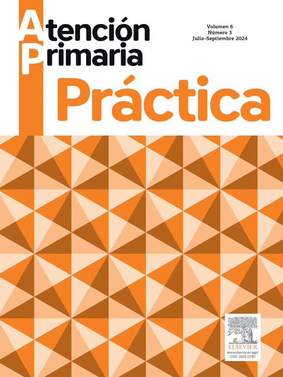
Case presentationA 70-year-old autonomous portuguese male with arterial hypertension and dyslipidemia. This patient self-referred to the Acute Disease Care Consultation (ADCC) at his Primary Care Center (PCC) with a 3-day history of periumbilical pain, without clear relief or worsening factors. The patient reported periumbilical swelling and purulent discharge, with localized infra-umbilical abdominal pain (Fig. 1). He denied other symptoms such as fever, chills, sweating, nausea, vomiting, other gastrointestinal, or genitourinary concerns.

Physical examination revealed periumbilical swelling, erythema, and abdominal tenderness. Omphalitis was assumed as the initial clinical impression and the patient was empirically treated with oral co-amoxiclav 875 mg/125 mg twice a day and topic fusidic acid 20 mg/g for 7 days.
By the end of this initial treatment, there was no improvement. The patient was reassessed at the PCC and later referred for hospital evaluation by the General Surgery emergency department (ED) team. An ultrasound revealed no significant changes, and the patient was discharged with flucloxacillin 500 mg, 3 times a day for 16 days.
Two months later, despite local wound care and antibiotic treatment, no improvement was verified. The patient was again admitted to ED and repeated an abdominal ultrasound that showed a 24×15 mm heterogeneous liquid collection, with supraumbilical and paramedian topography, an inferior communicating path towards the umbilical region, and an estimated maximum thickness of 12 mm.
A computed tomography (CT) scan revealed 2 fluid and gas collections with skin communication: one of the collections with retroumbilical walls, measuring 26×29×42 mm, probably related to an infected US and another one, supraumbilical, in the subcutaneous cellular tissue, measuring 21×23×25 mm.
Disinfection, local anesthesia, and a 15 mm incision were performed. Purulent content drainage, exploration, and cleansing of the 60 mm depth collection cavity were conducted. The patient was discharged, continuing the prescribed antibiotics and was instructed to do wound dressing changes and disinfection at his PCC.
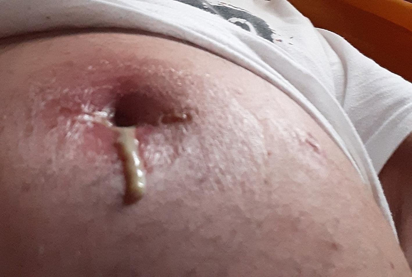
He was diagnosed with recurrent omphalitis and due to a patent urachus, underwent elective umbilical amputation surgery. The procedure included both umbilical herniorrhaphy and hernioplasty with a pedicled advancement flap to create a neoumbilicus. The patient achieved full recovery following the procedure and has not presented new omphalitis to date (Fig. 2). He maintains his regular appointments at the PCC.
Discussion
US is mostly found in male children and are rare in adults, making diagnosis difficult to achieve. Most cases are completely asymptomatic until complications arise. Both characteristics are present in this clinical case. Lower abdominal pain, fever, cutaneous inflammatory signs, a tender infraumbilical mass, and umbilical discharge are the main clinical manifestations of US, and all presented in this patient.
Failure in urachus' obliteration leads to other different types of abnormalities: patent urachus, in which the entire structure fails to close; urachal cyst, in which both ends close and there is an open central portion; and alternating sinus, draining either to the bladder or umbilicus.1 Other main differential diagnosis in adults include patent omphalomesenteric duct, omphalitis, complicated umbilical hernia, and Meckel's diverticulum. Ultrasonography is a good method as an initial screening. Other imaging scans such as CT, sinography, or magnetic resonance imaging (MRI) may be used. In this particular case, ultrasonography was not clear and CT scan allowed the diagnosis.3
The management of US depends on the presence of associated conditions or complications. If not treated promptly it can have an adverse outcome, such as fistulas, infection, and even sepsis development. As shown in this report, if no effective treatment is performed, complications such as peritonitis, riacho-colonic fistula, and neoplastic transformation may also arise. There is an ongoing debate regarding the optimal approach to treating infected US, which may involve a single-stage procedure (drainage and excision) with appropriate antibiotics, or a 2-stage process involving initial incision and drainage, followed by excision of the urachal remnant. In our case study, antibiotics were administered initially before excision was performed. For non-infected cases, radical 1-step excisions can be undertaken to remove the entire lesion, with or without bladder cuff removal, to mitigate potential malignancy-related complications that could arise from incomplete resection.3,4
ConclusionThis case underscores the significance of including infected US in the differential diagnosis of abdominal pain, one of the main reasons for patient admission to Acute Disease Care Consultation at PCCs. It also emphasizes that although rare, infected US is a potentially serious condition prompting timely diagnosis and intervention. Utilizing ultrasound or CT scan as complementary diagnostic tools can aid in accurate identification. Treatment typically involves antibiotic therapy and, given the potential for complications and malignant transformation, complete surgical excision is strongly recommended.
Primary care services are the National Healthcare Services' entrance and the main accountable for the systems' gatekeeping. As such, family physicians play an active role in acute disease care and management, besides having a deep knowledge of referral criteria for secondary care services. They also encourage treatment continuation and reassessment. These are essential points to maintain good recovery.
Hospital and PCC protocols on publication of patient data have been followed, the patient has provided full consent, and his privacy has been respected.
Ethical considerationsThe manuscript represents a case report for which the patient has provided full consent. The patient has signed and dated an informed consent form, authorizing the publication of his case and the presentation of the accompanying photo. The manuscript contains no identifying information. The authors have complied with all relevant ethical regulations. Furthermore, this work does not involve experimentation on animals or human subjects.
FundingThis work did not receive any specific grant from funding agencies in the public, commercial, or non-profit sectors.
