covid
Buscar en
Gaceta Mexicana de Oncología
Toda la web
Inicio Gaceta Mexicana de Oncología Use of Endocervical Brushing after Cervical Electrical Loop Excision as Predicto...
Información de la revista
Vol. 10. Núm. 5.
Páginas 265-268 (septiembre 2011)
Compartir
Compartir
Descargar PDF
Más opciones de artículo
Vol. 10. Núm. 5.
Páginas 265-268 (septiembre 2011)
Acceso a texto completo
Use of Endocervical Brushing after Cervical Electrical Loop Excision as Predictor of Residual Disease
Valor del cepillado de canal endocervical remanente durante LLETZ como predictor de enfermedad residual
Visitas
7033
Jorge Ojeda-Ortiza, Rebeca Muñoz-Molinaa, Francisco De la Rosa Bayónb, Beatriz Sereno Gómezc, Martha Guevara Cruzd
a Departamento de Colposcopia, Hospital General de Pachuca, Secretaría de Salud, Hidalgo, México. Supported by the Hospital General de Pachuca, Secretaría de Salud, Hidalgo, México.
b Departamento de Patología, Hospital General de Pachuca, Secretaría de Salud, Hidalgo, México. Supported by the Hospital General de Pachuca, Secretaría de Salud, Hidalgo, México.
c Academia Mexicana de Cito-patología, México. Supported by the Hospital General de Pachuca, Secretaría de Salud, Hidalgo, México.
d Instituto Nacional de Ciencias Médicas y Nutrición Salvador Zubirán, México, DF. Supported by the Hospital General de Pachuca, Secretaría de Salud, Hidalgo, México.
Este artículo ha recibido
Información del artículo
Resumen
Texto completo
Bibliografía
Descargar PDF
Estadísticas
Figuras (1)
Tablas (1)
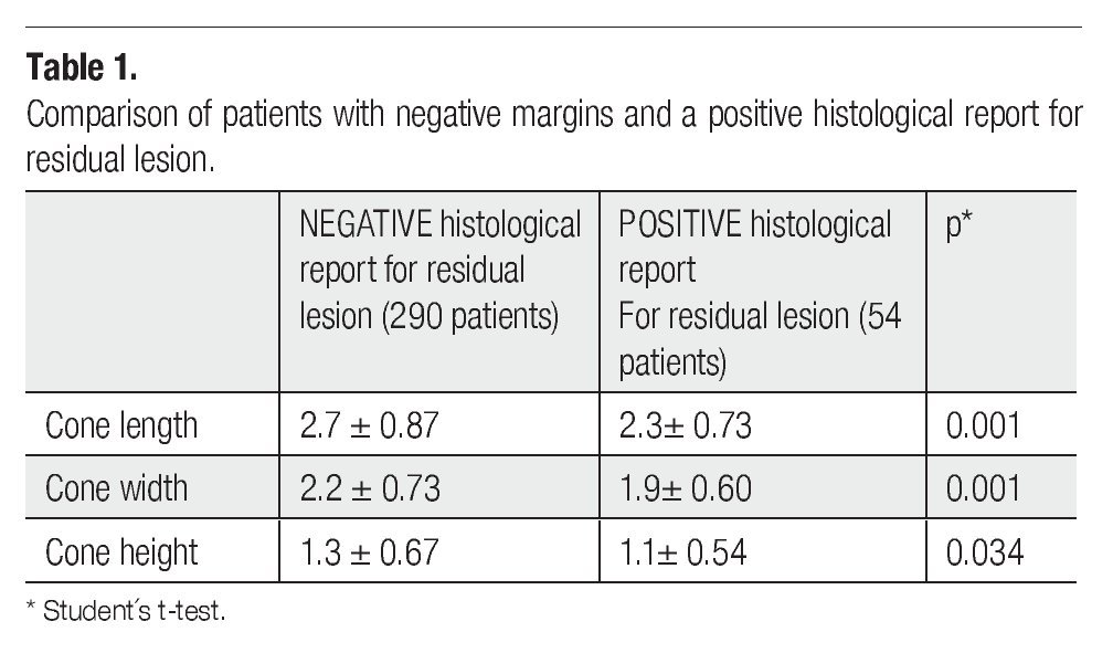
Table 1. Comparison of patients with negative margins and a positive histological report for residual lesion.
Objetivo: Hay varios factores que determinan la persistencia o recurrencia de la neoplasia intraepitelial cervical después de una escisión con asa grande de la zona de transformación (LLETZ). El presente estudio estima que el valor predictivo positivo del cepillado endocervical del canal cervical remanente inmediatamente después de retirar la muestra LLETZ, como elemento predictivo de enfermedad residual. Métodos: Estudio prospectivo, llevado a cabo desde enero de 1990 hasta diciembre de 2006, en el Hospital General de Pachuca, Hidalgo, México. LLETZ se realizó en 1817 pacientes con un concomitante cepillado cervical para estudio citológico. 344 pacientes tuvieron lesiones intraepiteliales de alto grado en la citología, histología y la colposcopia. En 62 pacientes con diagnóstico de lesiones intraepiteliales de alto grado tanto en LLETZ como en cytobrush, se realizó un segundo tratamiento en 6 ± 2 meses, ya fuese con LLETZ (16 pacientes) o histerectomía (54 pacientes). La evaluación histológica fue realizada por dos patólogos, cada uno desconocía el diagnóstico del otro patólogo (cegado). El análisis estadístico se realizó mediante t-test y prueba de ji cuadrada con el programa SPSS v10.0 (SPSS Inc., Chicago, IL). Resultados: Tras el segundo tratamiento de 62 pacientes, 87% (54 pacientes) tenían una lesión histológica probada residual y 13% (8 pacientes) no tenían enfermedad residual; estos resultados tuvieron significación estadística (p <0 05 conclusiones: cuando los márgenes endocervical y el cepillado cervical son positivos de forma concomitante existe un alto valor predictivo 87 tener la enfermedad residual según se muestra en segundo espécimen
Palabras clave:
LLETZ, enfermedad residual, predictivo, margen endocervical, México
Objective: There are several factors which determine the persistence or recurrence of cervical intraepithelial neoplasia after a large-loop excision of the transformation zone (LLETZ). The current study estimates the positive predictive value of endocervical brushing of the remaining cervical canal immediately after removing the LLETZ specimen, as a predictive element of a residual disease. Methods: Prospective study carried out from January 1990 to December 2006, at General Hospital of Pachuca, Hidalgo, Mexico. LLETZ was performed in 1817 patients undergoing concomitant cervical brushing for cytological study; 344 patients had a high grade intraepithelial lesion on cytology, histology, and colposcopy. In 62 patients with diagnosis of high grade intraepithelial lesion in both LLETZ and cytobrush, a second treatment was performed within 6 ± 2 months either with LLETZ (16 patients) or hysterectomy (54 patients). Histological evaluation was performed by two pathologist, each blinded to the diagnosis of a pathologist. Statistical analyses were performed by t-test and2 test using SPSS v10.0 (SPSS Inc., Chicago, IL). Results: Following the second treatment performed in 62 patients, 87% (54 patients) had a histological proven residual lesion and 13% (8 patients) had no residual disease; these results had a statistical significance of p <0 05 conclusions: when endocervical margins and cervical brushing are concomitantly positive there is a high predictive value 87 of having residual disease as demonstrated in second specimen
Keywords:
LLETZ, residual disease, predictive, endocervical margin, Mexico
Texto completo

The contributions of the authors were as follows: JO: conception, design, and supervision of the study; RM: general design, medical examination, manuscript preparation, and data interpretation; FJDB: Pathologist to study the biopsies; BS: contributed to the experimental design; MG: general design, statistical analysis, and manuscript preparation. All authors agreed on the final version of the manuscript. Authors had no conflict of interest.

¿ INTRODUCTION

Electrical excision of cervical lesions was first used in the nineteenth century. Hunner (1906) was the first reporting the use of therapeutic electrocauterization.1 Rene Cartier in 1981 adapted a small loop electrode using low voltage and in 1989 Walter Prendiville for the first time introduced radio frequencies to perform the excision of the cervical transformation zone using a large loop (LLETZ).2,3 This technique has become widely used and offers the advantages of obtaining a large specimen for histopathological diagnosis, it is well tolerated by patients when performed under local anesthesia on an outpatient basis, has few complications and side effects, and has a relatively low cost;4 in addition, it allows to correlate colposcopic impression with the histology, and micro-invasion can be ruled-out.5 Also, fertility is preserved. Disadvantages include post-treatment cervical stenosis,6 infections, bleeding,7 and thermal damage of the margins that may preclude an adequate study of the margins.4

LLETZ is an effective treatment of cervical intraepithelial neoplasia (CIN) and thus avoids the progression to invasive carcinoma.

The persistence or recurrence of CIN following LLETZ is related to several factors such as degree of the disease,8 positive endocervical curettage,9 diagnosis of positive margins of the specimen.9-11 Ghaem-Maghami demonstrated that after incomplete resection, the relative risk (RR) of having a recurrence of disease of any degree was 5.47 (confidence interval [CI] 95% 4.37-6.83).12 Tan et al. report that only positive margins after LLETZ are an independent risk factor (p <0.05, odds ratio [OR] = 4.20)13 and Livasy comments on the presence of involved endocervical glands as a factor.14

Chien-Hsing points out the usefulness of clinical indicators such as positive endocervical margins, age over 50, and disease in multiple quadrants as having a positive predictive value of 40%, 31.4% and 21.9%, respectively.15 The management of patients with residual lesions post LLETZ is still controversial. Incomplete resection is ignored on many occasions when patients are followed. Follow-up is also done by cytology, a second loop excision, and even hysterectomy. There are no reports in the literature on endocervical brushing of the cervix immediately post LLETZ with histological study of the specimen vs. endocervical brushing in cases with positive margins as a predictor of residual disease.

¿ OBJECTIVES

To study the concordance of positive predictive value of endocervical brushing performed immediately following LLETZ and histological results of cervical cones with positive endocervical margins, as predictors of residual disease.

¿ METHODS

A prospective study including 1817 patients referred with abnormal cytology and treated by LEETZ was undertaken at the Colposcopy Clinic, General Hospital of Pachuca, Hidalgo, Mexico.

The Ethics and Research Committee from the same hospital approved the protocol; informed consent was obtained from all patients.

Demographic data included age, educational level, marital status; reproductive data included number of pregnancies and children, age at sexual debut, number of sexual partners, and method of contraception; clinical data included previous cytological results, index cytology, Reid index,16 and Campion's level.17

These 1877 patients underwent endocervical brushing of the remaining endocervical canal immediately after removing the specimen; 344 patients had a high grade intraepithelial lesion on cytology, histology, and colposcopy. In 62 patients margins were positive on histology and the endocervical brushing was also positive. These 62 patients underwent a new treatment (LEETZ or hysterectomy) within 6 (± 2) months.

All 1877 patients were referred to the Colposcopy Clinic due to an abnormal cytology. On the first visit, colposcopy was performed and recorded using the modified Reid Colposcopic Index. Colposcopy-directed biopsies were obtained and sent for histo-pathological studies. On a second visit, a LLETZ was performed under local anesthesia. Each specimen was marked at 12 o'clock and reported by a single Pathologist. A second expert pathologist reviewed all 62 cases blinded to the previous results. An excellent concordance between both pathologists was reported (kappa = 0.95). All margins were considered acceptable for evaluation.

Statistical analysis was performed on SPSS for Windows (version 10.00; SPSS Inc. Chicago, Ill.). Continuous variables were expressed as means and standard deviation; dichotomy variables as frequencies and percentages. To study group comparisons student´s t test for independent samples . A significant value was expressed using a two-tail p value <0.05.

¿ RESULTS

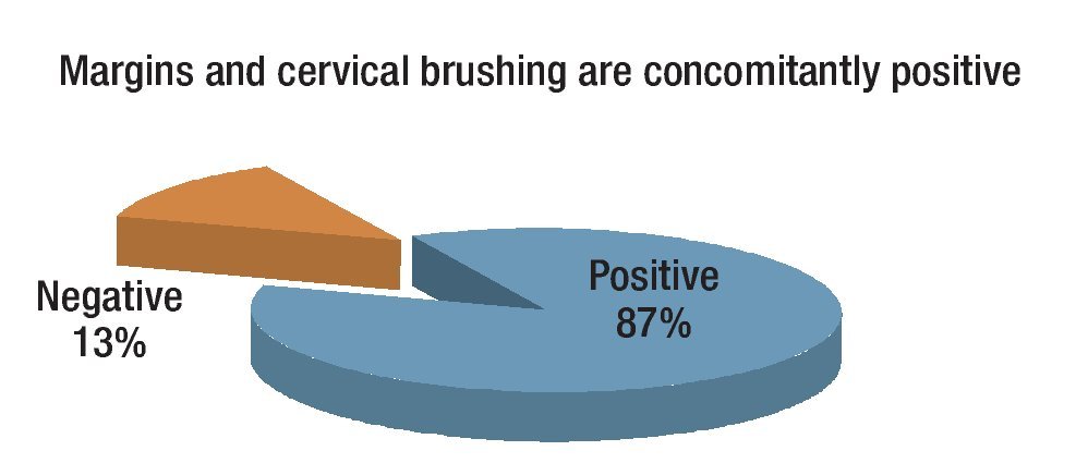
Mean age of the 344 patients with cytological and histological diagnosis of high grade intraepithelial lesion was 42.2 ± 12.5 years (range 20 - 80). The 62 cases with both positive endocervical margins and a positive cytobrush of the remaining canal had a second treatment: 46 hysterectomies and 16 second LLETZ. Findings showed 54 patients (87%) had residual lesion on the second specimen and 8 (13%) had a negative specimen (p <0.05) (Figure 1).

Figure 1. Results in 62 second specimens with both positive endocervical margins and endocervical brushing.

The length, height and width of the cone were significantly different in patients with residual disease when compared with those from patients with negative margins and negative endocervical brushing (Table 1).

There were no significant differences in demographic data, reproductive history, and clinical data between the group of patients with residual disease and the one with both negative margins and endocervical brushing.

¿ DISCUSSION

When the presence of a positive endocervical cytobrush and endocervical margins positive in a cone (LLETZ) specimen were combined, second specimens with residual lesion were obtained in 87% of the cases, i.e., such rate of positive specimens in the future may be predicted (positive predictive value). Residual lesion was seen in 8% of the cases (from 1817 LLETZ specimens). No other studies comparing Mexican rates have been published; however, rates reported in the international literature are quite variable (14% - 31%).18-21

¿ CONCLUSIONS

The positive predictive value of cone specimens with positive endocervical margins and concomitantly positive endocervical cytobrush of the proximal cervix is 87%. This knowledge may in the future allow choosing with reasonable certainty patients requiring a second treatment, and offering better results in the treatment of residual disease when taking into account these elegibility factors.

¿ ACKNOWLEDGMENT

To Fernando Guijon, MD for his commentaries on the manuscript and help in the translation.


Corresponding Author: Jorge Ojeda-Ortiz.

Departamento de Colpos-Departamento de Colposcopia, Hospital General de Pachuca - Carretera Pachuca-Tulancingo S/N Colonia Ciudad de los niños. Pachuca, Hidalgo. 42070, México.

Phone and Fax (52) 77 1718 2500.

Email:jorgeojeda@hotmail.com

Bibliography
[1]
Wright TC, Richart RM, Ferenczy A..
Electrosurgery for HPV-related diseases of the lower genital tract..
A practical handbook for diagnosis and treatment by loop electrosurgical excision and fulguration procedures. ,
[2]
Prendiville W, Cullimore J, Norman S..
Large-loop excision of the transformation zone (LLETZ)..
A new method of management for woman with cervical intraepithelial neoplasia. , 96 (Br J Obstet Gynecol 1989), pp. 1054-60
[3]
Prendiville W..
Large loop excision of the transformation zone..
, 38 (Clin Obst Gynecol 1995), pp. 622-27
[4]
Montz FJ, Holschneider CH, Thompson DR..
Large loop excision of the transformation zone..
[5]
Trejo O, Hurtado H, de la Torre FE, Ojeda J, Tamariz E, López JL..
Atlas de patología cervical, México, 1999..
[6]
Wright TC, Richart RM, Ferenczy A..
Electrosurgery for HPV-related diseases of the lower genital tract, New York, NY, Arthur Vision, 1991..
[7]
Ferenczy A, Choukround D, Arseneau J..
Loop electrosurgical excision procedure for squamous intraepithelial lesions of the cervix: advantages and potential pitfalls..
, 87 (Obstet Gynecol 1996), pp. 332-37
[8]
Hulman G, Pickles CJ, Gie CA, et al..
Frequency of cervical intraepithelial neoplasia following large loop excision of the transformation zone..
, 51 (J Clin Pathol 1998), pp. 375-77
[9]
Felix JC, Muderspach LI, Duggan BD, Roman LD..
The significance of positive margins in loop electrosurgical cone biopsies..
, 84 (Obstet Gynecol 1994), pp. 996-1000
[10]
Mohamed-Noor K, Quinn MA, Tan J..
Outcomes after cervical cold knife conization with complete and incomplete excision of abnormal epithelium: a review of 699 cases..
, 67 (Gynecol Oncol 1997), pp. 34-38
[11]
Kobak WH, Roman LD, Felix JC, et al..
The role of endocervical curettage at cervical conization for high-grade dysplasia..
, 85 (Obstet Gynecol 1995), pp. 197-201
[12]
Ghaem-Maghami S, Sagi S, Majeed G, Soutter WP..
Incomplete excision of cervical intraepithelial neoplasia and risk of treatment failure: a meta-analysis..
, 8 (Lancet Oncol 2007), pp. 985-93
[13]
Tan XJ, Wu M, Lang JH, et al..
Predictors of residual lesion in cervix after conization in patients with cervical intraepithelial neoplasia and microinvasive cervical cancer..
, 89 (Zhonghua Yi Xue Za Zhi 2009), pp. 17-20
[14]
Livasy CA, Maygarden SJ, Rajaratnam CT, Novotny DB..
Predictors of recurrent dysplasia after a cervical loop electrocautery excision procedure for CIN-3: a study of margin, endocervical gland and quadrant involvement..
, 12 (Mod Pathol 1999), pp. 233-8
[15]
Lu CH, Liu FS, Kuo CJ, et al..
Prediction of persistence or recurrence after conization for cervical intraepithelial neoplasia III..
, 107 (Obstet Gynecol 2006), pp. 830-35
[16]
Reid R, Stanhope CR, Herschman BR, et al..
Genital warts and cervical cancer. IV..
A colposcopic index for differentiating subclinical papillomaviral infection from cervical intraepithelial neoplasia. , 149 (Am J Obstet Gynecol 1984), pp. 815-23
[17]
Campion M, Ferris D, Guijón F..
Colposcopia moderna: Un enfoque práctico. A.S.C..
C. , (P Primera Edición en Español 1995),
[18]
Socolov D, Anton G, Anton AC, et al..
Electrosurgical loop excision/conisation for cervical intraepithelial neoplasia in an algorithm that excludes punch biopsy--a study of 210 cases..
Chirurgia (Bucur). , 104 (2009), pp. 295-301
[19]
Paraskevaidis E, Kalantaridou SN, Paschopoulos M, et al..
Factors affecting outcome after incomplete excision of cervical intraepithelial neoplasia..
, 24 (Eur J Gynaecol Oncol 2003), pp. 541-43
[20]
Ghaem-Maghami S, Sagi S, Majeed G, Soutter WP..
Incomplete excision of cervical intraepithelial neoplasia and risk of treatment failure: a meta-analysis..
Lancet Oncol. , 8 (2007), pp. 985-93
[21]
Hulman G, Pickles CJ, Gie CA, et al..
Frequency of cervical intraepithelial neoplasia following large loop excision of the transformation zone..
, 51 (J Clin Pathol 1998), pp. 375-77
Descargar PDF
Opciones de artículo