


Providing high-quality myocardial perfusion myocardial single-photon emission computed tomography (SPECT) images while keeping the radiation dose at a low level for patients with cardiovascular disease is a challenging issue in nuclear medicine. In this research, optimization of imaging protocol by using the idea of irregular imaging was proposed as a potential technique to achieve this goal.
MethodsHumanoid digital phantoms for male and female in 3 sizes of big, medium, and small were produced in this study. We examined 2 sets of imaging protocols on these phantoms. The standard regular imaging protocol includes 32 views at equiangular distances (5.6°). The irregular imaging protocol is implemented by increasing the number of views at anterior to left lateral angular span while decreasing the views at other angles. We compared the image quality of both protocols in terms of contrast, resolution, and signal-to-noise ratio (SNR).
ResultsThe images resolution at 4 walls of the heart (Septal, Lateral, Inferior, Anterior) were evaluated for both proposed and conventional methods and showed that the proposed irregular imaging protocol resulted in images with enhances resolution at Septal, Lateral, and Inferior images. The wall to background contrast and signal-to-noise ratio for the short-axis cross-sections were also calculated and showed the superiority of irregular imaging protocol over the conventional approach.
ConclusionThe results presented in this study suggest that irregular imaging protocol can be a promising approach in nuclear medicine for patients with cardiovascular disease.
Proporcionar imágenes de tomografía computarizada por emisión de fotón único (SPECT) miocárdicas de perfusión miocárdica de alta calidad mientras se mantiene la dosis de radiación en un nivel bajo para los pacientes con enfermedades cardiovasculares es un tema desafiante en la medicina nuclear. En esta investigación, se propuso la optimización del protocolo de imágenes utilizando la idea de imágenes irregulares como una técnica potencial para lograr este objetivo.
MétodosEn este estudio se produjeron fantasmas digitales humanoides para hombres y mujeres en tres tamaños: grande, mediano y pequeño. Examinamos dos conjuntos de protocolos de imágenes en estos fantasmas. El protocolo estándar de imágenes regulares incluye 32 vistas a distancias equiangulares (5,6°). El protocolo de imágenes irregulares se implementa aumentando el número de vistas en el intervalo angular anterior al lateral izquierdo mientras se reducen las vistas en otros ángulos. Comparamos la calidad de imagen de ambos protocolos en términos de contraste, resolución y SNR.
ResultadosLa resolución de las imágenes en las cuatro paredes del corazón (septal, lateral, inferior, anterior) se evaluó tanto para los métodos propuestos como para los convencionales y mostró que el protocolo de imagen irregular propuesto resultó en imágenes con resolución mejorada en las imágenes septal, lateral e inferior. También se calcularon el contraste de la pared y el fondo y la relación señal-ruido para las secciones transversales del eje corto y mostraron la superioridad del protocolo de imagen irregular sobre el enfoque convencional.
ConclusiónLos resultados presentados en este estudio sugieren que el protocolo de imagen irregular puede ser un enfoque prometedor en medicina nuclear para pacientes con enfermedades cardiovasculares.
Myocardial perfusion imaging (MPI) using single-photon emission computed tomography (SPECT) is widely used to diagnose ischemic heart disease. At present, approximately 9 million myocardial perfusion imaging studies are performed annually in the United States and 250 000 studies in Japan including perfusion, fatty acid imaging, and sympathetic nerve imaging.1
SPECT is generally recognized as a low-resolution imaging modality. Conventional SPECT images acquired using rotating Anger cameras have an average resolution of 15 mm under clinical conditions, and thus the quality is inferior to that of high-resolution images acquired using magnetic resonance imaging and X-ray computed tomography.2 However, given that perfusion is an initial evaluation by nuclear cardiology, incomplete SPECT images may be acceptable in the era of multidimensional cardiac imaging.
Due to the limited number of counting, attenuation, or scattering of photons by various factors such as noise, SPECT image quality may be damaged.3 Perhaps, low level of injected radioactive tracer is the most important inherent limitation of nuclear cardiology confirming the concerns raised by high radiation load, the importance of trying to keep the exposure to the lowest possible radiation level, and the development of methods to reduce radiation dose for patients with cardiovascular disease (CVD), and also avoiding increased dose to enhance image quality. Another way to reduce this problem is by increasing imaging time. However, long acquisition times make imaging sensitive to patients̓ motion in motor artifacts.4
Thus, a particular consideration of the details of both imaging and image processing protocols is required to ensure high detection quality in the myocardial perfusion studies.5 Recently, sampling theory plays an essential role in signal processing.6 In this way, many papers published in the last decades have investigated some traditional methods in the angular sampling field,7–9 whereas several novel algorithms have been developed in the past decade.10 Ultimately, based on these conducted studies, both national and international professional societies have developed some practical guidelines for the nuclear laboratories to maintain the high-quality nuclear services.11
It should be noted that due to the position of the heart in the left hemithorax, an angular sampling span is defined as a standard circuit with a 180° angular sampling from right anterior oblique (RAO) (45°) view to left posterior oblique (LPO) (45°) view. Meanwhile, the optimal number of projections depends on the number of projections matching the system resolution. Furthermore, to avoid a resolution reduction and for higher quality images using Tc99m and HR collimator, at least 60–64 projections via regular angular spans for the 180° circuit are required to be considered.12
Here, as the irregular sampling comes from some signal processing issues, conducting such researches has more focused on the irregular signal sampling as well as its reconstruction in the past.13,14 In this matter, Hans et al. proposed some studies. They investigated the irregular reconstruction based on the pure mathematics theory.13–15 Besides, a reconstruction of the irregular samples is evaluated based on Fourier mathematics.6,16 It is worthwhile to mention that as the image is really a two-dimensional signal, it is possible to generalize and extend the irregular reconstruction signal issues to the reconstruction of the tomographic images. As such, the idea of irregular sampling to enhance the quality of the reconstructed images seems to be applied without any cropping more views. The purpose of this study is to increase the number of views (decrease the sampling angle) in the angles where we have the most view of the heart and also reduce the number of views (increase the sampling angle) in the angles where the view of the heart is limited due to the ribs.
This increased at angles where we have a better view of the heart is expected to result in the collection of a higher proportion of primary photons than scattered photons that cause background events and reduced image quality, and essentially problematic for all nuclear medicine imaging, especially SPECT.
Therefore, in this paper, we obtained the cross-sectional images using irregular sampling (inconsistent angles between views), and then compared their qualities with the traditional method.
MethodsThe human phantomIn this study, SPECT images of the myocardial were obtained using the NCAT digital human phantom. The original phantom model was established from CT data using Non-uniform rational B-spline (NURBS) cardiac and torso (NCAT) phantom that are capable of producing anatomical maps of the body with the desired resolution and size. This simplified torso model has up to 20 materials consisting of myocardium and pericardium, blood pool, ventricles, arteries, veins, muscle, gallbladder, lungs, spleen, stomach, rib bone, cortical bone, spine bone, and liver.
In this investigation, the male and female phantoms were produced with heights of 176 and 165 cm, respectively, in 3 sizes of big, medium, and small by using BMI standard with a resolution of 1.56 mm (6 phantoms in total). Activity (Tc99m) in the heart (target organ) and adjacent organs (background radiation) was determined based on the results of clinical studies.
Imaging protocolMonte Carlo (MC) simulations were performed using SimSET code, version 2.6.2.6 for SPECT camera (GE Infinia Hawkeye, USA). Imaging was performed for each phantom at a 180-degree orbit around the patient's body in 2 stages, one using the standard protocol (32 views with an equal distance of 5.6°) and the other using the proposed protocol. At the angular span of RAO (45°) to anterior, with 10° angular distance, 5 views were performed. On the other hand, at the angular span of anterior to left lateral with 4 degrees angular distance, 22 views were carried out. Besides, at the angular span of the left lateral to LPO (45°), with 10° angular distances, 5 views were performed. Totally, the same 32 views are acquired in the standard imaging protocol.
The diffusion information was achieved with a matrix 64 × 64 pixels and a 20% energy window to the center of 140 keV. In the end, all the views obtained by FBP were reconstructed. Afterward, the Butterworth filter parameters were applied according to the values utilized in the clinic (cut-off = 0.44, order = 0.8), and finally, the dataset was evaluated.
Image quality parametersFunctional parameters including spatial resolution, contrast, and signal-to-noise ratio were evaluated for both protocols.
ResolutionThe resolution of the images was calculated in the 4 walls of the heart (Septal, Lateral, Inferior, Anterior) at short-axis sections by plotting a profile along with 2 perpendicular directions on the left ventricular image in different slices. The count vs. location curve was plotted, and the degree of flattening in the curves at half-maximum height was calculated as an estimate of resolution.4
ContrastThe organ contrast of the NACT phantom reconstructed images was calculated by Eq. (1).
where A and B represent the average count in the myocardial wall and the extra-cardiac area, respectively.16
Locations of the left intraventricular muscles in each slice were determined in the images reconstructed from Short-Axis (SA) sections. Then, the ratio of the mean count in the left ventricular muscle area to mean count in intraventricular muscle in the test images was considered as the contrast of each image. Computations were done automatically using MATLAB code to avoid human errors.4
Signal-to-noise ratio (SNR)The definition of the SNR is the mean value of the perfusion values in the mid regions of the myocardium normalized to the standard deviation of cavity that was calculated by Eq. ((2):

Given that photon count follows the Poisson statistical distribution, one can obtain:

Therefore, ratio of root mean square to mean represents SNR.4,17
ResultsResolutionTable 1 demonstrates resolution values (in pixels) for the 4 walls of the heart (Septal, Lateral, Inferior, Anterior) at short-axis sections of the proposed method and the conventional method. An improvement was observed in the resolution of the proposed method (irregular sampling), especially for obese-sized phantoms.
Resolution values in the 2 conventional and proposed methods, which compare the 2 methods, show a satisfactory improvement in the resolution obtained by the proposed method.
| Sex | Regular | Irregular | ||||||
|---|---|---|---|---|---|---|---|---|
| Lateral | Anterior | Septal | Inferior | Lateral | Anterior | Septal | Inferior | |
| Male | ||||||||
| Big | 11.3301 | 12.1051 | 10.2535 | 11.2173 | 9.39342 | 12.1184 | 9.42775 | 10.6868 |
| Medium | 11.8124 | 12.7903 | 10.1483 | 11.5077 | 9.76374 | 12.279 | 9.35797 | 11.2911 |
| Small | 12.7792 | 13.4898 | 11.2995 | 12.6843 | 10.8117 | 13.5537 | 9.84444 | 12.0899 |
| Female | ||||||||
| Big | 14.4932 | 13.2151 | 11.7507 | 12.5178 | 11.0496 | 12.5303 | 10.1873 | 11.676 |
| Medium | 15.5507 | 13.2707 | 12.314 | 12.546 | 11.9109 | 13.1974 | 10.2817 | 11.9676 |
| Small | 16.4911 | 14.1143 | 15.2857 | 14.8386 | 13.6292 | 14.4632 | 11.3934 | 13.1699 |
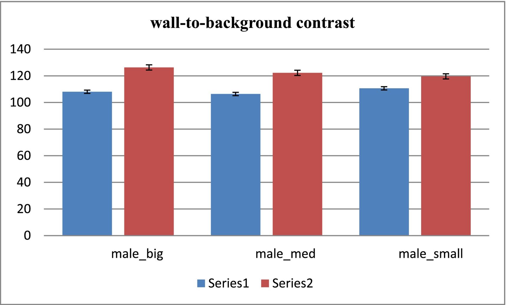
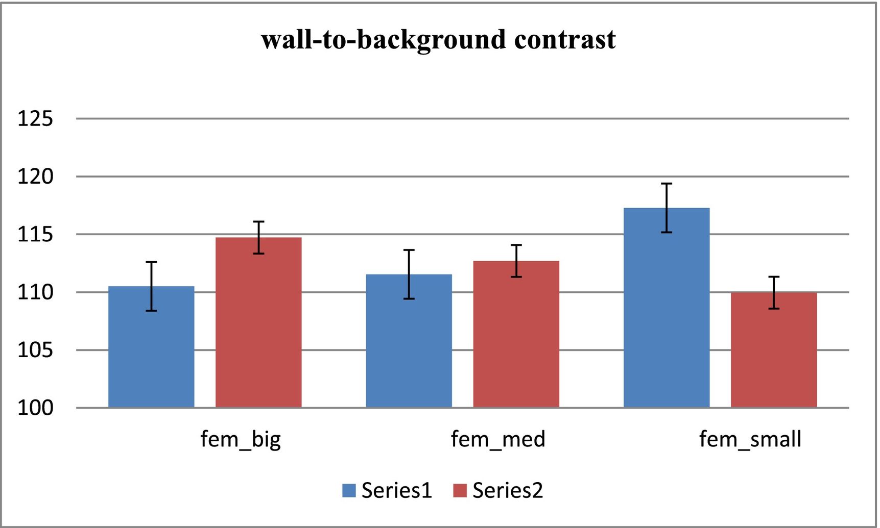
After reconstructing the short-axis images, the wall-to-background contrast was calculated for all 6 phantoms. The results for the contrast of the male and female phantoms are shown in Figs. 1 and 2, respectively. Improvement of the contrast was observed for the proposed method (irregular sampling), except for the case of the small phantom of the female.

.")
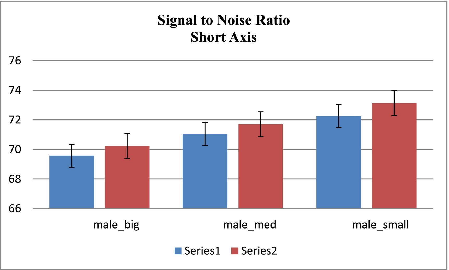
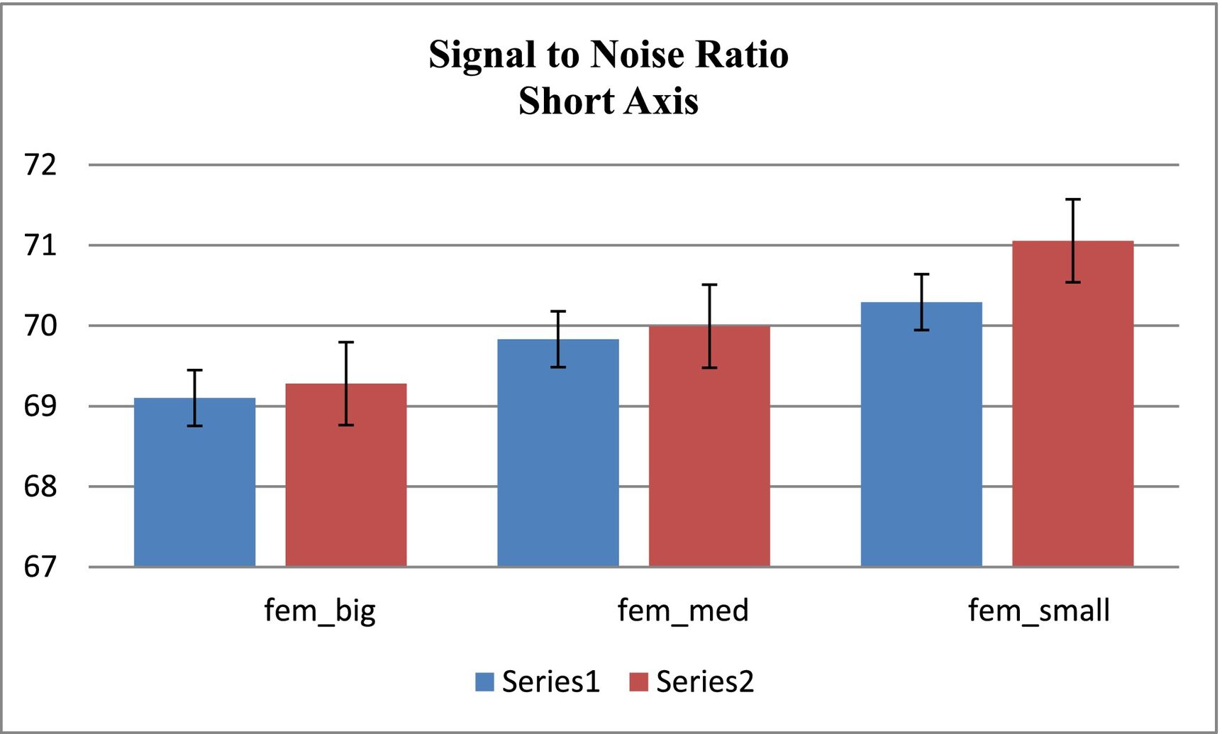
As mentioned earlier, the signal-to-noise ratio for the short-axis cross-section was calculated by using MATLAB code. The results of these calculations for the male and female phantoms are shown in Figs. 3 and 4, respectively. The results are indicating an improvement in the signal-to-noise ratio in the proposed method in comparison to the conventional method.


In this study, to improve image quality, we proposed an irregular sampling method inspired by signal processing. The image quality parameters in the proposed method were calculated and compared with the conventional method. The results of these calculations confirm the quality improvement in the proposed method. The gamma radiations used in nuclear medicine are constituted by photons and characterized by their energy, which inversely is proportional to their wavelength. The gamma rays come from the nuclei during the nuclear reactions and are mono energetics for a given characteristic reaction.18 Gamma photons contain much higher energy than visible light and can pass out of the body.19
There are 3 factors affecting photon attenuation. First is energy, which is the characteristic of the radiation itself. The photon of higher energy is not easier to be attenuated and is easier to penetrate materials. The other 2 factors are related to materials; named density and atomic number, respectively. The probability of interacting with photons increases when the larger values of density and atomic number.20 Despite the optimal energy of gamma rays (140 keV), the beams emitted from the heart are attenuated due to the anatomical position of the heart and the clinical imaging of cardiac SPECT is mainly limited by photon statistics.21 As the heart is asymmetrically and angularly enclosed between ribs on the left side of the chest, the gamma rays used in Tc99m imaging have to travel 15–20 cm from the tissue to reach the camera. By assuming an average linear attenuation coefficient of 0.18 cm, only 3–7 percent of the photons may reach the camera without any attenuation.22 The interpretation and quantification of myocardial perfusion SPECT are hampered by photon attenuation, Compton scatter, and collimator resolution effects. These factors preclude a linear relationship between the counts in the image and the true tracer distribution.23
Since the accuracy of reconstructed images is directly related to the effects of resolution and damping on projection data, the spatial resolution is not substantially improved by SPECT scanning.24 In this way, the presence of scattered beams in the SPECT images is one of the critical factors to reduce the contrast and resolution of the image and causing errors in quantitative computing.25 The scattered rays that are well known as the background noise, maybe larger than useful information several times. However, in this study, we improved the resolution, contrast, and signal-to-noise ratio of the heart walls by decreasing the attenuated photons associated with breast in women and the diaphragm in men23 and by increasing the initial photon count.
Finally, regarding the great importance of improving the image quality parameters in the irregular sampling, this method can be considered as a good modality for the standard protocol, especially in the cases where the weakening of the breast and diaphragm restricts the detection of the walls. Thus, by becoming SPECT as an important part of nuclear medicine, more increasing of the image contrast is expected to better distinguish the small lesions as well as to define a better edge of large lesions, and generally better visual interpretation of the scans. Therefore, it is suggested to investigate the image quality by the proposed method and by placing the defect in the heart wall.
FundingThere is no funding to declare.
Ethical approvalThere is no human or animal subjects and the study is exempt from ethical approval.
Authors contributionMR: study design, performing and writing; NZ: study concept, data collection, analysis and drafting. Both authors read and approved the study.
There is no acknowledgment for the present study.
