Los hemangiomas congénitos (HC) son tumores vasculares benignos presentes al nacimiento. Se describen 2 tipos: RICH (rápidamente involutivo) y NICH (no involucionan y crecen de forma proporcional al niño). La regresión de algunos HC es incompleta, estos se denominan PICH (parcialmente involutivos).
ObjetivoDescribir una serie de 14 casos de HC asistidos en nuestro centro entre 2016-2019, destacando sus características clínico-evolutivas.
MetodologíaEstudio observacional, descriptivo retrospectivo.
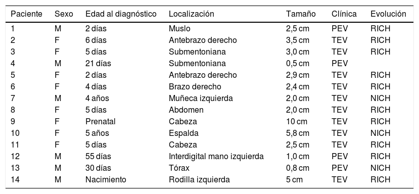
ResultadosEncontramos en nuestra serie una incidencia similar en ambos sexos, la edad media al momento de la consulta fue 15,8 días, en 2 de los pacientes el diagnóstico fue retrospectivo. En un caso el diagnóstico fue prenatal. La forma más frecuente de presentación fue la tumoración eritematoviolácea con telangiectasias y palidez periférica, seguido de la placa eritematoviolácea. La localización más frecuente fue en miembros superiores, seguido de la cabeza, el dorso alto, el tórax, el muslo, el abdomen y el miembro inferior. Diez casos fueron catalogados como RICH, 3 como NICH, y un paciente no continuó en seguimiento. Se realizó una ecografía Doppler a todos los pacientes que se confirmó el diagnóstico. Ninguno de los pacientes presentó complicaciones. Se realizó seguimiento clínico cercano, no requiriendo intervenciones.
ConclusionesEl diagnóstico de HC es clínico-ecográfico. El tratamiento está indicado cuando existen complicaciones, siendo el seguimiento clínico lo más aconsejado.
Congenital hemangiomas (CH) are benign vascular tumors present at birth. Two types of CH are identified: RICH (rapidly involuting cogenital hemangioma) and NICH (noninvoluting, grows proportionally with the child). Regression of some CH is incomplete; these are known as PICH (partially involuting).
ObjectiveTo describe a series of 14 cases of CH treated in our unit during the period 2016-2019, focusing on its clinical-evolutionary features.
MethodologyObservational, descriptive retrospective study of cases of CH treated in a reference clinic.
ResultsIn our case-series we found a similar incidence in both sexes, the mean age at the time of consult was 15.8 days, in two of the patients the diagnosis was retrospective. In one case, the diagnosis was prenatal. The most frequent form of presentation was an erythematous-violaceous tumor with telangiectasias and peripheral pallor. Followed by the erythematous-violaceous plaque. The most frequent location was in the upper limbs, followed by the head, upper back, thorax, thigh, abdomen and lower limb. Ten cases were classified as RICH, 3 cases as NICH, and in one case the patient did not continue to be followed up. Doppler ultrasound was performed on all patients that confirmed the diagnosis. None of the patients presented complications. Close clinical follow-up was performed, requiring no interventions.
ConclusionsDiagnosis of CH is clinical-echographic. Treatment is indicated when there are complications, with clinical follow-up being the most recommended course of action.
Artículo
Comprando el artículo el PDF del mismo podrá ser descargado
Precio 19,34 €
Comprar ahora


