El tamaño del cuerpo calloso (CC) se ha relacionado con la presencia de déficits cognitivos y emocionales en diversos trastornos neuropsiquiátricos y del estado de ánimo. Dado que estos déficits se observan también en la conducta suicida, hemos investigado específicamente la asociación entre la atrofia del CC y dicha conducta.
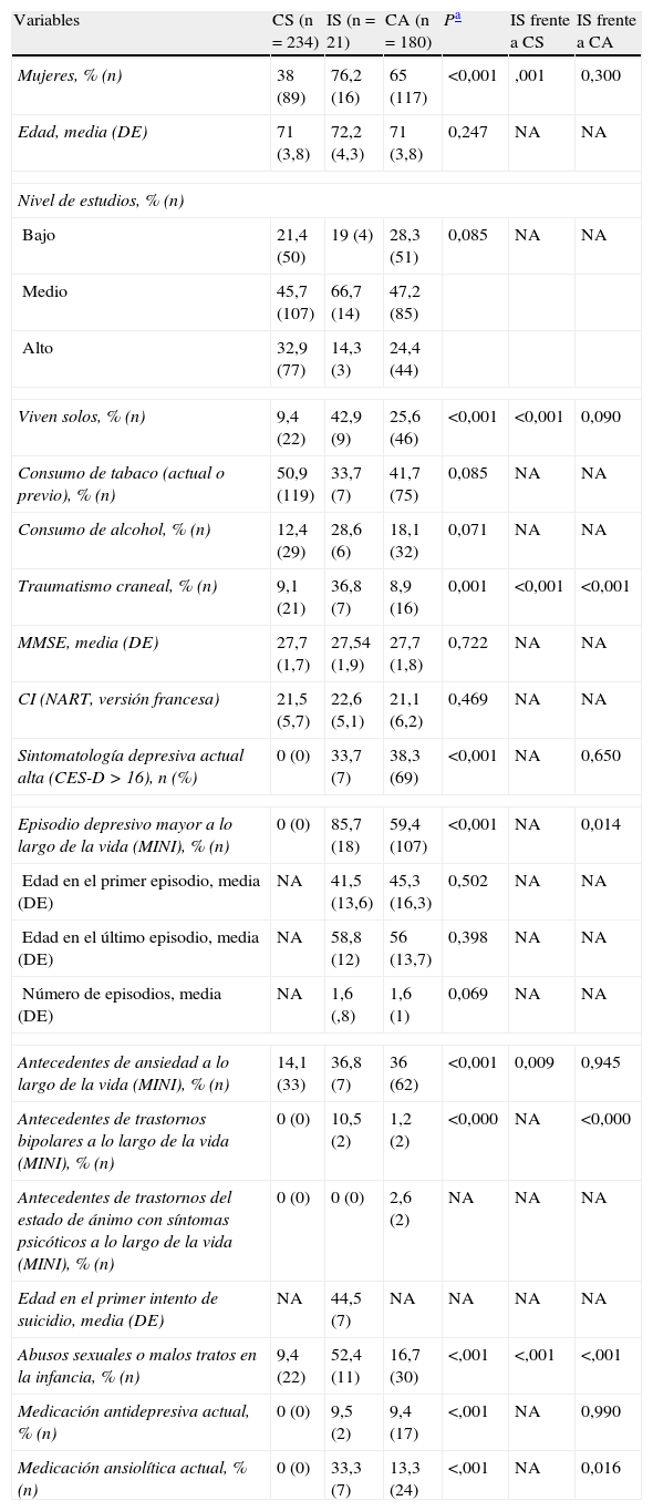
MétodosEstudiamos a 435 individuos diestros, sin demencia, de una cohorte de base comunitaria formada por personas de edad igual o superior a 65 años (estudio ESPRIT). Dividimos a los participantes en 3 grupos: individuos con intentos de suicidio (n=21), individuos de control afectivos (CA) (n=180) sin antecedentes de intentos de suicidio pero con antecedentes de depresión, e individuos de control sanos (CS) (n=234). Se utilizaron imágenes de resonancia magnética con ponderación T1 para medir las áreas mesosagitales del CC anterior, medio y posterior. Se aplicó un análisis de covarianza multivariado para comparar las áreas del CC de los 3 grupos.
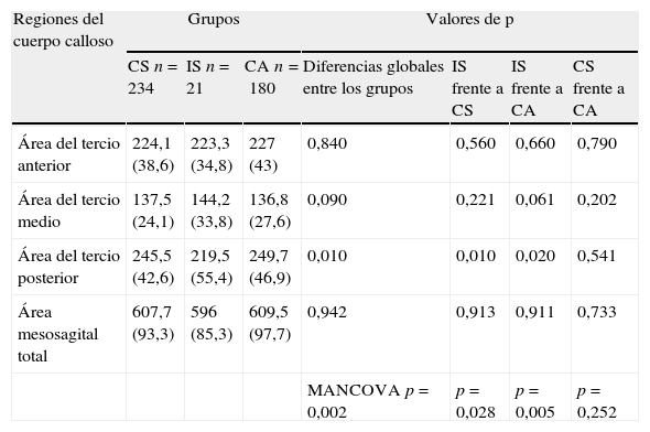
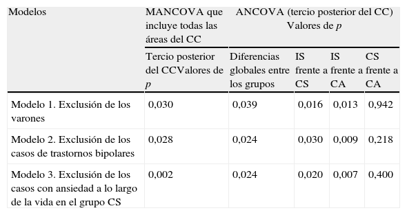
ResultadosLos análisis multivariados, con un ajuste en cuanto a edad, sexo, trauma infantil, traumatismo craneal y volumen encefálico total, mostraron que el área del tercio posterior del CC era significativamente menor en los individuos con intentos de suicidio, en comparación con los CA (p=0,020) y los CS (p=0,010). No se observaron diferencias significativas entre CA y CS. No hubo diferencias en cuanto a los tercios anterior y medio del CC.
ConclusionesNuestros resultados resaltan la presencia de un tamaño reducido del tercio posterior del CC en los individuos con antecedentes de suicidio, lo cual sugiere una disminución de la conectividad interhemisférica y un posible papel del CC en la fisiopatología de la conducta suicida. Serán necesarios nuevos estudios para confirmar estos resultados y esclarecer las alteraciones celulares subyacentes que conducen a estas diferencias morfométricas.
Corpus callosum (CC) size has been associated with cognitive and emotional deficits in a range of neuropsychiatric andmood disorders. As such deficits are also found in suicidal behavior, we investigated specifically the association between CC atrophy and suicidal behavior.
MethodsWe studied 435 right-handed individuals without dementia from a cohort of community-dwelling persons aged 65 years and over (the ESPRIT study). They were divided in three groups: suicide attempters (n=21), affective control subjects (AC) (n=180) without history of suicide attempt but with a history of depression, and healthy control subjects (HC) (n=234). T1-weighted magnetic resonance images were traced to measure the midsagittal areas of the anterior, mid, and posterior CC. Multivariate analysis of covariance was used to compare CC areas in the 3 groups.
ResultsMultivariate analyses adjusted for age, gender, childhood trauma, head trauma, and total brain volume showed that the area of the posterior third of CC was significantly smaller in suicide attempters than in AC (P=.020) and HC (P=.010) individuals. No significant differences were found between AC and HC. No differences were found for the anterior and mid thirds of the CC.
ConclusionsOur findings emphasize a reduced size of the posterior third of the CC in subjects with a history of suicide, suggesting a diminished interhemispheric connectivity and a possible role of CC in the pathophysiology of suicidal behavior. Further studies are needed to strengthen these results and clarify the underlying cellular changes leading to these morphometric differences.
Artículo
Comprando el artículo el PDF del mismo podrá ser descargado
Precio 19,34 €
Comprar ahora


