Evaluar las características radiológicas e histológicas de los cánceres de intervalo (CI) y de cribado diagnosticados durante el periodo 2007-2018, en un total de 6 rondas de un programa poblacional de cáncer de mama.
Material y métodosEl estudio incluye las mujeres con edades entre 50 y 69años a las que se realizó una mamografía digital con intervalo bienal y lectura simple. Se diagnosticaron un total de 1.395 carcinomas de cribado y 300CI durante ese periodo.
Para la clasificación de los CI se realizó un proceso de revisión retrospectivo (lecturas ciega e informada) al final de cada ronda y se registraron los hallazgos radiológicos, la densidad mamaria, las características histológicas, el fenotipo y el tratamiento quirúrgico.
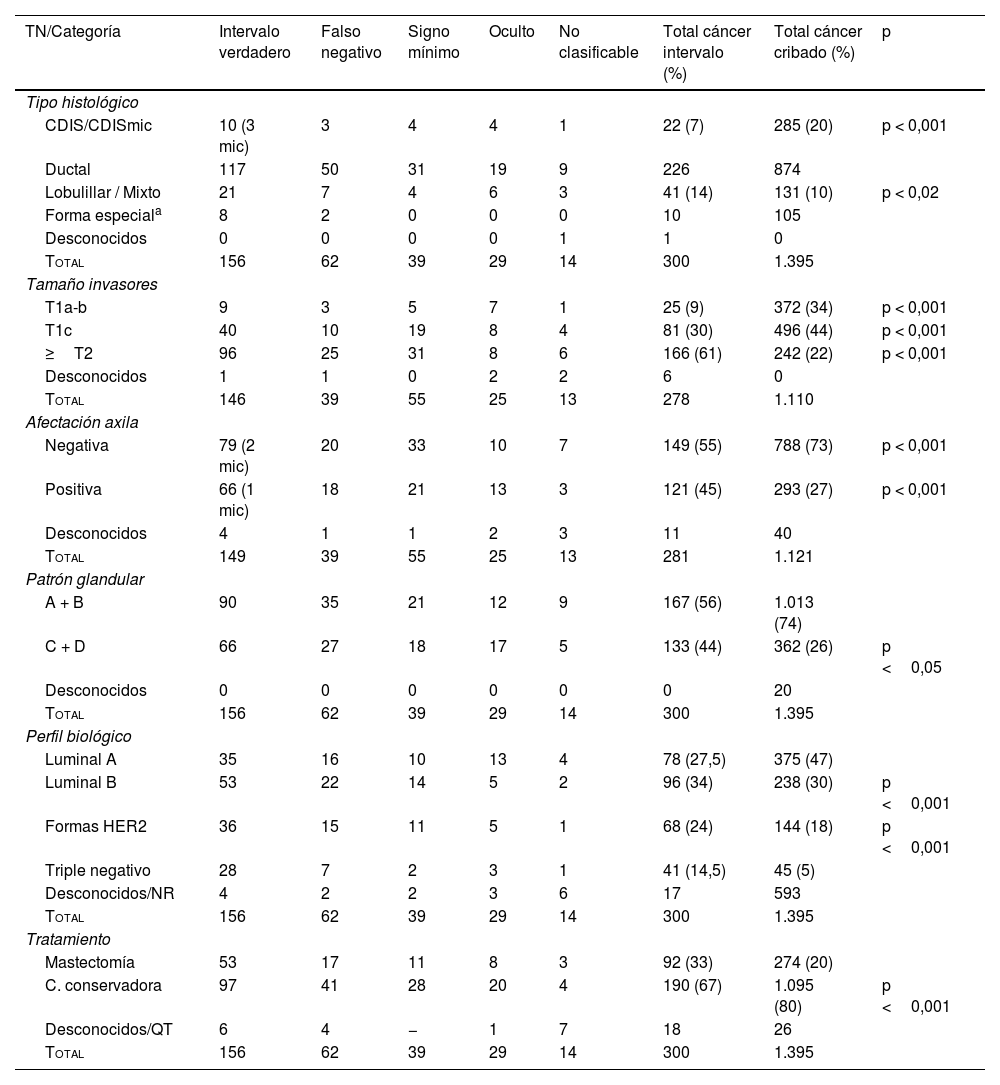
ResultadosLa clasificación de los CI fue: 156 (52%) intervalo verdadero, 62 (20,5%) falso negativo, 39 (13%) signo mínimo, 29 (9,5%) oculto y 14 (5%) inclasificable.
Retrospectivamente, los hallazgos radiológicos más frecuentes fueron: masa/asimetría (64%), calcificaciones (16%), distorsión (13%) en los casos de falso negativo, y masa/asimetría (58%) y calcificaciones (31%) en los casos de signo mínimo.
Respecto a las características histopatológicas, hubo diferencias significativas en los cánceres infiltrantes tipo T1a-b (9% de los CI vs 34% de los del cribado; p<0,001); T1c (30% de los CI vs 44% del cribado; p<0,001); T2 o mayor (61% de los CI vs 22% de los del cribado; p<0,001) y grado de afectación axilar (45% de los casos de CI vs 27% de los del cribado; p<0,001).
Respecto a los casos con subtipos más agresivos (HER2+ y triple negativo), también se apreciaron diferencias: 38,5% en los CI vs 23% en los cánceres de cribado; p<0,001.
Se confirmó un significativo menor número de mastectomías (p<0,001) en los carcinomas del cribado (67%) en comparación con las realizadas en los CI (80%).
ConclusionesLos CI pueden ser atribuibles a la actividad del radiólogo hasta en el 20% de los casos. El hallazgo radiológico más frecuente es la asimetría/masa y presentan un estadio más avanzado al diagnóstico que los cánceres de cribado, por lo que son tratados de una forma más radical. La revisión de los CI debe formar parte del control de calidad de un programa de cribado.
To analyze the radiologic and histologic characteristics of screening and interval cancers diagnosed in the period comprising 2007 through 2018 in a total of six rounds of a population-based breast cancer screening program.
Material and methodsWe analyzed 1395 carcinomas detected at screening and 300 interval carcinomas diagnosed in women aged 50 to 69years old who underwent digital mammography every two years during the study period. Screening mammograms were read once.
To classify the interval carcinomas, we retrospectively reviewed (blind reading followed by unblinded reading) at the end of each round, recording the radiologic findings, breast density, histologic characteristics, phenotype, and surgical treatment.
ResultsThe interval carcinomas were classified as true interval cancers in 156 (52%) cases, false-negatives in 62 (20.5%), minimal signs in 39 (13%), occult lesions in 29 (9.5%), and impossible to classify in 14 (5%).
Retrospectively, the most common radiologic findings in the false-negative cases were mass/asymmetry (64%), calcifications (16%), and distortion (13%); the most common radiologic findings in the cases with minimal signs were mass/asymmetry (58%) and calcifications (31%).
There were significant differences in the histologic characteristics between cancers detected at screening and interval cancers: T1a-b (9% of the interval cancers vs. 34% of those detected at screening; P<.001); T1c (30% of the interval cancers vs. 44% of those detected at screening; P<.001); T2 or greater (61% of the interval cancers vs. 22% of those detected at screening; P<.001), and the degree of axillary involvement (45% of the interval cancers vs. 27% of those detected at screening; P<.001).
There were also significant differences between cancers detected at screening and interval cancers in the proportion of cases with more aggressive subtypes (HER2+ and triple-negative): 38.5% of the interval cancers vs. 23% of those detected at screening; P<.001.
A significantly higher proportion of interval cancers were treated with mastectomies (80% vs. 67% of those detected at screening; P<.001).
ConclusionsAbout 20% of interval cancers were evident on screening mammograms. The most common radiologic finding in interval cancers was asymmetry/mass. Interval cancers are diagnosed at a more advanced stage than cancers identified at screening, so they are more often treated by mastectomy. Reviewing interval cancers is essential for quality control in screening programs.
Artículo
Comprando el artículo el PDF del mismo podrá ser descargado
Precio 19,34 €
Comprar ahora


