Analizar el efecto de la edad, el sexo y el valor b en el coeficiente de difusión aparente (CDA) de áreas cerebrales alteradas en las enfermedades neurodegenerativas.
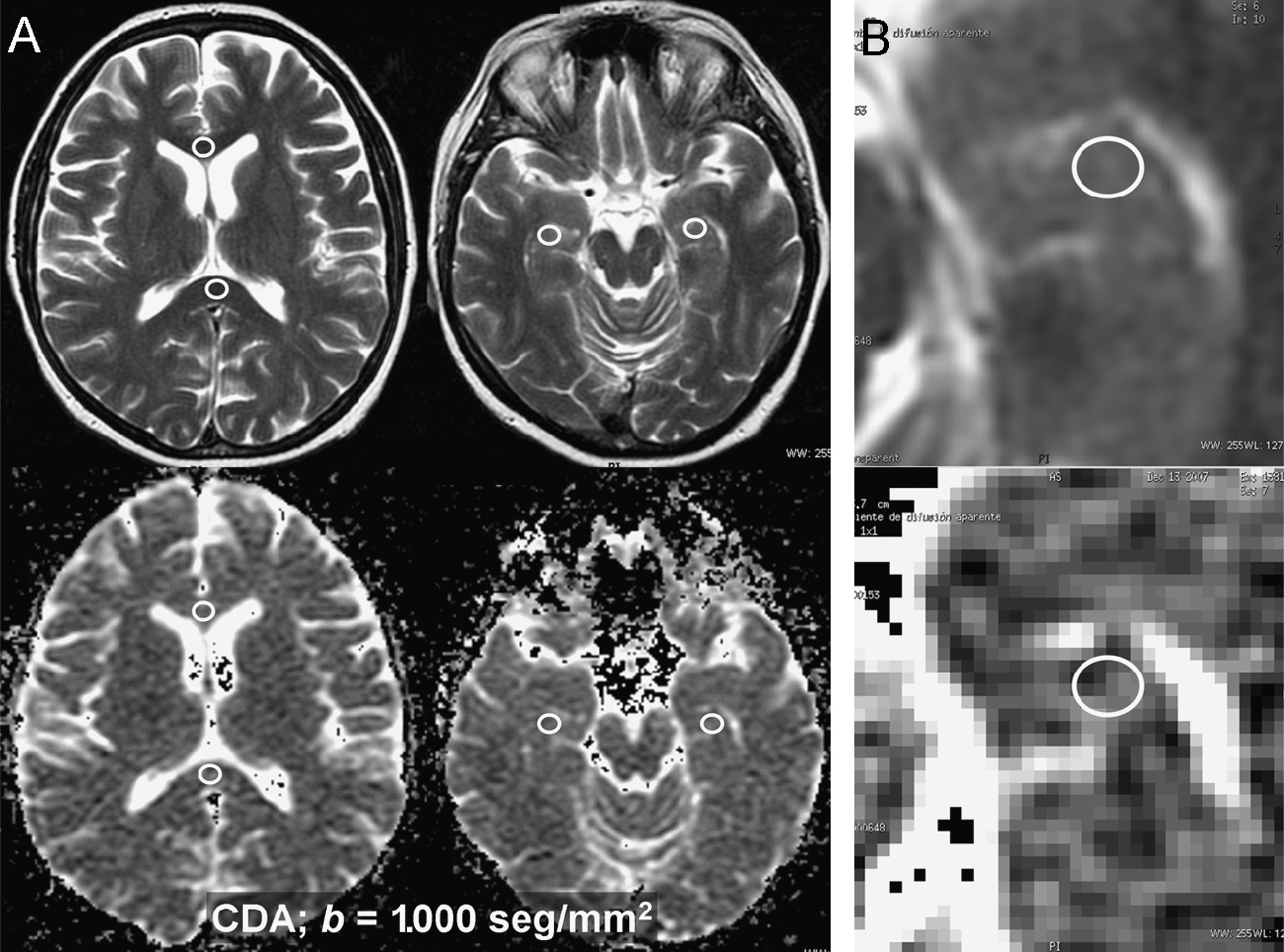
Material y métodosSe estudió el CDA de hipocampo, rodilla y esplenio del cuerpo calloso en 50 pacientes normales, con difusión por resonancia magnética b1.000 y b3.000s/mm2. Para cada localización se calcularon las diferencias entre los CDA b1.000 y b3.000 (diferencial de difusión [DD]). Se separaron los pacientes en grupos de edad (⩽30, 31–60 y >60 años). El análisis de las diferencias de CDA y DD entre grupos de edad y entre sexos se hizo con los tests de ANOVA y Kruskal-Wallis, y la corrección de Bonferroni. Para la correlación del CDA y el DD con la edad se utilizó el test de Pearson.
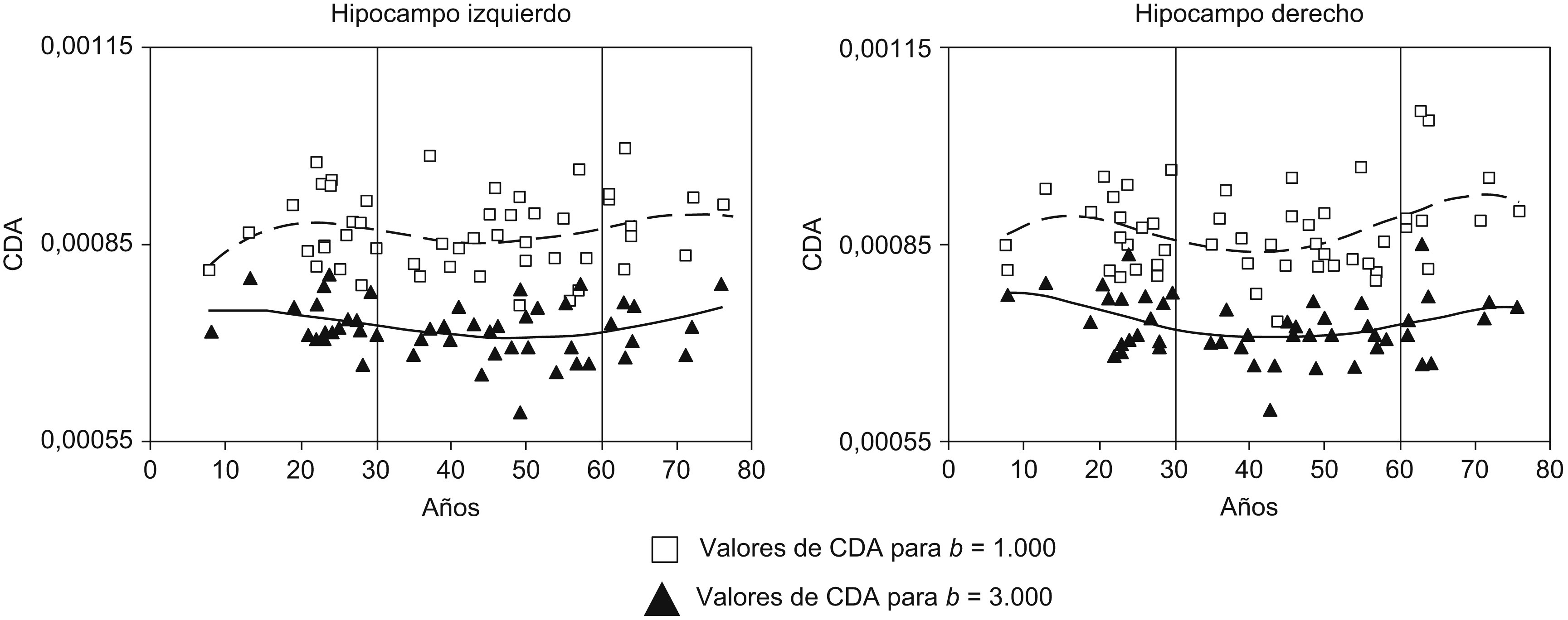
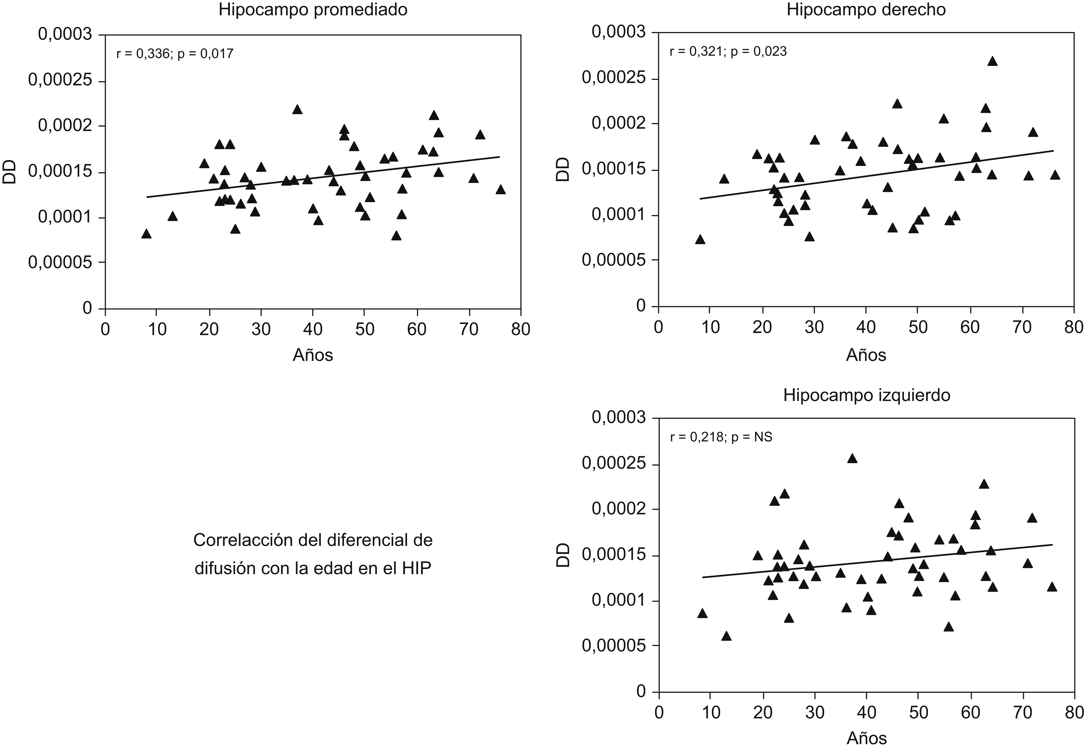
ResultadosEn el hipocampo derecho hubo diferencias de CDA (b1.000, p=0,011; b3.000, p=0,024) y DD (p=0,006) con la edad. Las diferencias del CDA se observaron entre los grupos de 31–60 años y >60 (p=0,009) para b 1.000, y entre<30 y 31–60 años (p=0,036) para b 3.000. El DD varió entre los >60 años y el resto. En el cuerpo calloso hubo diferencias significativas entre sexos en el DD de la rodilla (p=0,016). El DD mostró correlación con la edad en el hipocampo derecho (r=0,321, p=0,023).
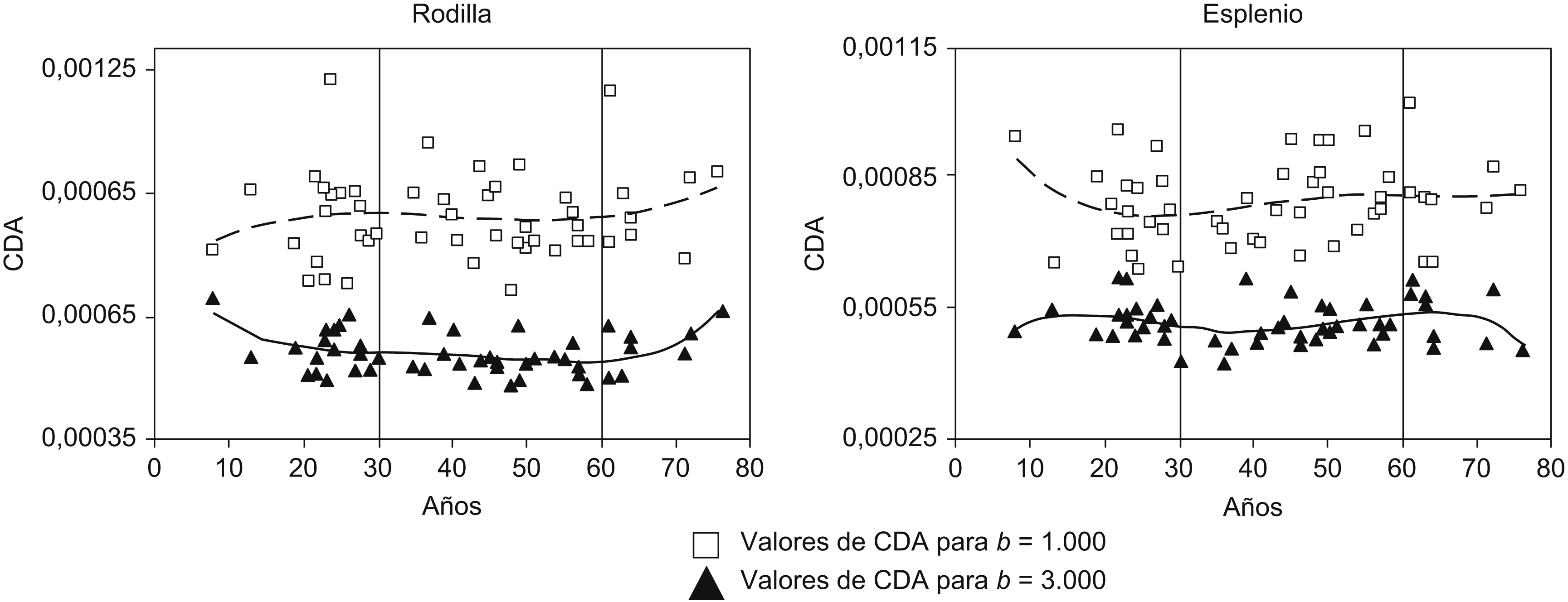
ConclusionesNuestros datos indican mayor estabilidad de los valores de CDA medidos con b3.000 durante el envejecimiento. El análisis del CDA con valor b más alto puede ser de utilidad en las enfermedades neurodegenerativas.
To analyze the effects of age, sex, and b value on the apparent diffusion coefficient (ADC) in brain areas affected by neurodegenerative diseases.
Material and methodsWe studied the ADC of the genu and splenium of the corpus callosum and of the hippocampus in normal patients using diffusion magnetic resonance imaging (dMRI) with b1,000s/mm2 and b3,000s/mm2. We calculated the differences between the ADC (diffusion differential [DD]) with b1,000 and with b3,000 for each region. Patients were classified into the following age groups (⩽30 years old, 31–60 years old, >60 years old). We used a Kruskal-Wallis one-way ANOVA and the Bonferroni correction to analyze the differences in ADC and DD between age groups and between sexes. Pearson's chi-square test was used to correlate the ADC and DD with age.
ResultsIn the right hippocampus, we observed differences in ADC (b1,000, p=0.011; b3,000, p=0.024) and DD (p=0.006) with age. Differences in ADC were observed between the 31–60 year-old age group and the >60 year-old age group (p=0.009) for b1,000, and between the<30 year-old age group and the 31–60 year-old age group (p=0.036) for b3,000. The DD in the >60 year-old age group was different from the rest. In the corpus callosum, there were significant differences between sexes in the DD of the genu (p=0.016). The DD was correlated with age in the right hippocampus (r=0.321, p=0.023).
ConclusionsOur data indicate greater stability in mean ADC values with b3000 during aging. It might be useful to analyze the ADC with a higher b in patients with neurodegenerative diseases.


